Блог

  • Omega-3 Index

    Omega-3 Index

    The role of the Omega-3 index (EPA + DHA in red blood cells) in the body

    The omega-3 index is a measure of the percentage of two key fatty acids (eicosapentaenoic EPA and docosahexaenoic DHA) in the membranes of red blood cells. This is an important marker of long-term Omega-3 intake and its impact on health.

    EPA and DHA functions:

    1. Cardiovascular system:
    • Reduce the level of triglycerides.
    • Reduce the risk of arrhythmias and atherosclerosis.
    • Maintain the elasticity of blood vessels.
    1. Brain and nervous system:
    • DHA is the main structural component of the brain and retina.
    • Improve cognitive function, reduce the risk of depression and dementia.
    1. Anti-inflammatory effect:
    • EPA suppresses chronic inflammation (reduces the level of IL-6, TNF-α).
    • Helps with autoimmune diseases (rheumatoid arthritis).
    1. Pregnancy and child development:
    • They are critical for the formation of the fetal brain and vision.
    • Reduce the risk of premature birth.

    Symptoms of Omega-3 Deficiency

    Lack of EPA and DHA is associated with an increased risk of chronic diseases.

    Possible signs:

    • Dry skin, eczema, brittle nails.
    • Fatigue, poor concentration, depression.
    • Frequent inflammation (such as joint pain).
    • Elevated triglyceride levels.
    • Visual impairment (reduced adaptation to darkness).

    Risk groups:

    • People who rarely eat fish (salmon, mackerel, sardines).
    • Vegans and vegetarians (if they don’t take seaweed supplements).
    • In diseases of the gastrointestinal tract (impaired fat absorption).

    Symptoms of an Omega-3 excess

    Overdose is possible only when taking high doses of supplements (more than 3 g / day).

    Side effects:

    • Blood thinning (risk of bleeding, especially when taking anticoagulants).
    • Nausea, diarrhea, bad taste in the mouth.
    • Decreased immune response (rarely, at very high doses).

    Important! An excess of Omega-3s from food (fish) is almost impossible.


    The norm of the Omega-3 index in the blood test

    The omega-3 index is measured in % of the total amount of fatty acids in red blood cells:

    • < 4%Deficiency (high risk of cardiovascular diseases).
    • 4-8%Average level (typical for most people).
    • > 8%Optimal level (associated with minimal risk of heart disease).

    Ideal target indicator: 8-12% (especially for the prevention of atherosclerosis).


    How to increase the Omega-3 index?

    1. Food:
    • Oily fish: salmon, herring, sardines (2-3 servings per week).
    • Seafood: shrimp, oysters.
    • Seaweed (a source of DHA for vegans).
    1. Supplements:
    • Fish oil (contains EPA + DHA).
    • Krill oil (better absorbed).
    • Seaweed oil (suitable for vegetarians).

    Dosage to maintain the index > 8%:

    • 500-1000 mg of EPA + DHA per day -for general health.
    • 2000-4000 mg / day – for high triglycerides or inflammation (only under the supervision of a doctor).

    Conclusion

    The omega-3 index is an important marker that reflects the balance of healthy fatty acids in the body.

    • The norm: 8-12% (red blood cell count).
    • Deficiency (<4%) increases the risk of heart disease, depression, and inflammation.
    • An excess is only possible if you overdose on supplements (risk of bleeding).

    To maintain your health, include fatty fish in your diet or take Omega-3 capsules, especially if you are at risk.

  • Antibodies to thyroglobulin-AT-TG

    Antibodies to thyroglobulin-AT-TG

    Thyroglobulin antibodies (AT-TG): role, symptoms of imbalance, and norms

    1. The role of AT-TG in the body

    Thyroglobulin (TG) is a precursor protein for thyroid hormones (T3 and T4), and AT– TG is an autoantibody that attacks this protein. Their presence indicates autoimmune processes in the thyroid gland.

    Thyroglobulin functions:

    • Storage of iodine for the synthesis of T3 and T4.
    • Participation in the production of thyroid hormones.

    Why determine AT-TG?
    These antibodies are a marker of autoimmune thyroid damage. , and their level increases when:

    • Autoimmune thyroiditis (Hashimoto’s disease).
    • Diffuse toxic goiter (Graves ‘ disease).
    • Thyroid cancer (monitoring after removal of the gland).

    2. Symptoms of excess AT-TG (autoimmune thyroiditis)

    A high level of AT-TG indicates the destruction of thyroid tissue, which leads to:

    • Hypothyroidism (if the gland is damaged):
    • Weakness, fatigue.
    • Weight gain, swelling.
    • Dry skin, hair loss.
    • Depression, constipation.
    • Hyperthyroidism (at the beginning of the disease, with a temporary release of hormones):
    • Irritability, anxiety.
    • Weight loss, sweating.
    • Rapid heartbeat.
    • Enlargement of the thyroid gland (goiter).

    3. Symptoms of AT-TG deficiency

    A low level or lack of AT-TG is normal, but if there are symptoms of thyroid dysfunction, the cause may be:

    • Non-autoimmune hypothyroidism (iodine deficiency, congenital disorders).
    • Subacute thyroiditis (viral inflammation).
    • Taking thyroid hormones (artificial suppression of TG).

    4. Norms of AT-TG in the blood test

    ConditionReference values (IU / ml)Interpretation
    Standard0-18 (depends on the lab)No autoimmune reaction
    Borderline18–50The initial stage of the autoimmune process is possible
    Increased value> 50Autoimmune thyroiditis, Graves ‘ disease

    Notes:

    • In 5-10% of healthy people , a moderately elevated level without pathology is possible.
    • In women over 50 years of age, AT-TG is often elevated without clinical manifestations.

    5. When is the test scheduled?

    • Diagnosis of autoimmune thyroiditis.
    • Control of Graves ‘ disease.
    • Monitoring of thyroid cancer recurrence (after surgery).
    • Examination for infertility or miscarriages (if an autoimmune process is suspected).

    6. Additional research

    • AT-TPO (antibodies to thyroperoxidase) is a more specific marker of autoimmune thyroiditis.
    • TSH, T3, T4 – assessment of thyroid function.
    • Ultrasound of the thyroid gland -detection of structural changes.

    7. What should I do in case of deviations?

    • If AT-TG is elevated, but the hormones are normal:
    • Follow-up with an endocrinologist 1-2 times a year.
    • TSH and ultrasound monitoring.
    • If you have hypothyroidism:
    • L-thyroxine replacement therapy.
    • If cancer is suspected:
    • Node biopsy, treatment by an oncologist.

    Important! AT-TG is not the cause of the disease, but a marker of the autoimmune process. .Thyroid dysfunction is treated not by antibodies, but by thyroid dysfunction.

  • Dehydrotestosterone (DHT)

    Dehydrotestosterone (DHT)

    The role of dehydrotestosterone (DHT) in the body

    Dehydrotestosterone (DHT) is an active metabolite of testosteroneproduced by the enzyme 5– alpha reductase. . It binds to androgen receptors 3-10 times more strongly than testosterone, and plays a key role in:

    1. Sexual development in men:
    • Formation of external genitalia in the fetus.
    • Growth of the prostate, seminal vesicles.
    • Development of secondary sexual characteristics (hair, voice).
    1. For women:
    • Regulation of pubic and armpit hair growth.
    • Affects the work of the sebaceous glands (in excess – acne, hirsutism).
    1. Hair and skin:
    • Control of beard and moustache growth in men.
    • In excess – androgenetic alopecia (male pattern baldness).
    1. The prostate:
    • Stimulates the growth of the gland (with hyperplasia-DHT accelerates the process).

    DHT tests

    The level of DHT is measured in the blood (less often-in tissues).

    Type of analysisStandards (depending on the laboratory)
    Men250-990 pg / ml (0.25-0.99 ng / ml)
    Women24-450 pg / ml (0.024-0.45 ng / ml)
    Children (up to puberty)< 50 pg / ml

    Note:

    • In men, DHT peaks in their 20s and 30s, then decreases.
    • In women, the level depends on the phase of the cycle (higher in the luteal phase).

    Symptoms of DHT deficiency

    A rare condition, most often associated with a genetic defect in 5-alpha reductase.

    For men:

    • Underdevelopment of the genitals (micropenis, hypospadias).
    • Weak growth of beard and moustache.
    • Lack of androgenetic alopecia (do not go bald).
    • Infertility (rare).

    For women:

    • Scanty pubic/underarmhair.
    • Dry skin (due to reduced sebaceous gland activity).

    Reasons:

    • Congenital 5-alpha reductase deficiency.
    • Taking 5-alpha reductase inhibitors (finasteride, dutasteride).

    Symptoms of excess DHT

    For men:

    • Androgenetic alopecia (receding hairline, baldness of the crown).
    • Prostate hyperplasia (frequent urination).
    • Aggressiveness, increased libido (rarely).

    For women:

    • Hirsutism (hair growth on the face, chest).
    • Acne, oily skin.
    • Androgenetic alopecia (thinning of hair on the temples).
    • Cycle disorders, infertility (with PCOS).

    Reasons:

    • Hyperandrogenism (PCOS, ovarian/adrenal tumors).
    • Taking anabolic steroids.
    • Idiopathic hypersensitivity of the receptors (even with normal DHT).

    How to normalize DHT levels?

    If there is an excess:

    1. 5-alpha reductase blockers:
    • Finasteride (Propecia) – reduces DHT by 60-70%.
    • Dutasteride (Avodart) – blocks both types of enzymes.
    1. Antiandrogens (for women):
    • Spironolactone, ciproterone acetate.
    1. Natural remedies:
    • Palmetto (Serenoa repens) is a weak inhibitor of 5-alpha reductase.
    • Curcumin, green tea (reduce inflammation).

    If there is a shortage:

    • In men – testosterone replacement therapy (partially converted to DHT).

    When is the test scheduled?

    1. Men:
    • Early baldness (up to 30 years).
    • Prostatic hyperplasia.
    1. Women:
    • Hirsutism, acne.
    • Suspected PCOS.

    Conclusion

    • DHT is a powerful androgen that affects hair, prostate, and sexual development.
    • Standards:
    • Men: 250-990 pg / ml.
    • Women: 24-450 pg / ml.
    • Deficiency is rare (in cases of genetic disorders).
    • Too much leads to baldness, hirsutism, and prostate problems.

    Example: If a woman has DHT &> 500 pg / ml + hirsutism – PCOS or adrenal tumor should be excluded.

  • Globulin

    Globulin

    Globulins: role in the body, symptoms of deficiency and excess, norms in the blood test

    Globulins are a group of plasma proteinsthat perform many functions, including immune defense, substance transport, and blood clotting. They are divided into several fractions determined by serum protein electrophoresis.


    1. Main types of globulins and their functions

    Globulin FractionKey representativesRole in the body
    Alpha-1 globulinsAlpha-1-antitrypsin, alpha-1-acid glycoproteinInhibit proteases, participate in inflammation
    Alpha-2 globulinsHaptoglobin, ceruloplasmin, alpha-2-macroglobulinThey bind hemoglobin, copper transport, and protease suppression
    Beta-globulinsTransferrin, hemopexin, complement components (C3, C4)Iron transport, participation in the immune response
    Gamma globulinsImmunoglobulins (IgG, IgA, IgM, IgE, IgD)Antibodies, protection against infections

    2. Symptoms of globulin deficiency

    Reduced globulin levels may be associated with:

    • Hypogammaglobulinemia (antibody deficiency → frequent infections).
    • Liver diseases (cirrhosis, hepatitis-protein synthesis disorders).
    • Nephrotic syndrome (loss of protein in the urine).
    • Genetic disorders (for example, alpha-1-antitrypsin deficiency).

    Symptoms:

    • Frequent bacterial and viral infections (with immunoglobulin deficiency).
    • Edema (due to a decrease in oncotic blood pressure).
    • Anemia (with transferrin deficiency-impaired iron transport).
    • Emphysema of the lungs (with alpha-1-antitrypsin deficiency).

    3. Symptoms of excess globulins

    An increase in the level of globulins is observed when:

    • Chronic inflammation and infections (tuberculosis, HIV, autoimmune diseases).
    • Multiple myeloma (a tumor that produces an excess of immunoglobulins).
    • Autoimmune diseases (rheumatoid arthritis, SLE).
    • Liver diseases (chronic hepatitis).

    Symptoms:

    • Increased blood viscosity (headaches, circulatory disorders).
    • Kidney damage (due to the deposition of abnormal proteins).
    • Enlargement of the lymph nodes and spleen (with myeloma, lymphomas).

    4. Norms of globulins in the blood test

    The reference values depend on the method of analysis (electrophoresis or biochemistry).

    A. Total globulin (biochemical analysis)

    GroupNorm (g / l)
    Adults20–35
    Children18–34

    B. Globulin fractions (electrophoresis)

    FractionNorm (% of total protein)Norm (g / l)
    Alpha-12–5%0,2–0,4
    Alpha-27–13%0,5–1,0
    Beta8–15%0,6–1,2
    Gamma12–22%0,8–1,6

    5. When is the test scheduled?

    • Suspected immunodeficiency.
    • Chronic infections or inflammation.
    • Diseases of the liver and kidneys.
    • Diagnosis of myeloma and other paraproteinemias.

    Important: Interpretation requires a comprehensive approach, taking into account other indicators (total protein, albumin, creatinine, liver tests).

  • C-peptide

    C-peptide

    C-peptide: role in the body, symptoms of deficiency and excess, norms in the blood test

    The role of C-peptide in the body

    C-peptide (connective peptide) is a fragment of the proinsulin moleculethat is broken off during the formation of insulin in the beta cells of the pancreas. .Its level reflects the insulin-producing ability of the pancreas.

    Main functions:

    • Marker of insulin synthesis (1 proinsulin molecule → 1 insulin molecule + 1 C-peptide molecule).
    • It helps to distinguish self-produced insulin from externally administered insulin(exogenous insulin does not contain C-peptide).
    • It is used to diagnose diabetes (especially if artificial hypoglycemia is suspected).
    • Evaluates the residual function of beta cells in patients with type 1 and type 2 diabetes.

    Symptoms of C-peptide deficiency

    A low level indicates insufficient insulinproduction, which occurs when:

    • Type 1 diabetes mellitus (autoimmune destruction of beta cells).
    • Long-term type 2 diabetes (depletion of pancreatic reserves).
    • Severe pancreatitis or removal of the pancreas.

    Symptoms:

    • Hyperglycemia (high blood sugar).
    • Thirst, frequent urination.
    • Weight loss, weakness (with type 1 diabetes).
    • Ketoacidosis (with severe insulin deficiency).

    Symptoms of excess C-peptide

    An elevated level indicates an excessive production of insulin, which happens when:

    • Insulin resistance(type 2 diabetes, obesity).
    • Insulinoma (a hormone-active pancreatic tumor).
    • Renal failure (C-peptide is excreted by the kidneys, with their dysfunction, its level increases).
    • Taking sulfonylureas (stimulate the release of insulin).

    Symptoms:

    • Hypoglycemia (low blood sugar): weakness, sweating, trembling, hunger.
    • Weight gain (due to hyperinsulinemia).
    • With insulinoma-attacks of hypoglycemia on an empty stomach.

    Blood levels of C-peptide

    Reference values depend on the analysis method and laboratory, but on average:

    ConditionNorm (ng / ml)Notes
    On an empty stomach0.8–4.2In healthy people
    After a mealUp to 6-8Peak in 1-2 hours
    Type 1 diabetes< 0.8Reduced due to beta cell destruction
    Type 2 diabetesIncreased or normalDepends on the stage of the disease
    Insulinoma> 4-5 on an empty stomachDiagnostic criteria

    Important:

    • The test is taken on an empty stomach (8-12 hours of hunger).
    • For differential diagnosis , tests are performed with glucose or fasting.
    • The level of C-peptide does not depend on the injected insulin (unlike the insulin itself).

    When is the test scheduled?

    1. Differentiation of type 1 and type 2 diabetes.
    2. Suspected insulinoma.
    3. Control of residual beta cell function.
    4. Suspicion of artificial hypoglycemia (secret use of insulin).
  • Osteocalcin

    Osteocalcin

    Osteocalcin: role in the body, symptoms of deficiency and excess, norms in the blood test

    The role of osteocalcin in the body

    Osteocalcin is a non-collagenic bone protein synthesized by osteoblasts (cells responsible for bone formation). It plays an important role in:

    • Bone mineralization -binds calcium and hydroxyapatites, helping to strengthen bone tissue.
    • Regulation of bone metabolism is a marker of bone formation (its level reflects the activity of osteoblasts).
    • Effects on carbohydrate and energy metabolism – osteocalcin is involved in the regulation of insulin secretion and tissue sensitivity to it.
    • Maintaining muscle function -affects muscle strength and endurance.

    Symptoms of osteocalcin deficiency

    Direct osteocalcin deficiency is rare, but a decrease in it may indicate:

    • Osteoporosis (especially in the elderly) – due to reduced bone formation.
    • Vitamin K deficiency (essential for osteocalcin activation).
    • Hypoparathyroidism (decreased function of the parathyroid glands).
    • Long-term use of glucocorticoids (inhibit osteoblasts).

    Symptoms:

    • Brittle bones, frequent fractures.
    • Muscle weakness.
    • Delayed healing of bone injuries.

    Symptoms of excess osteocalcin

    Elevated levels are usually associated with increased bone metabolism:

    • Paget’s disease (deforming osteitis).
    • Hyperparathyroidism (excess of parathyroid hormone).
    • Osteomalacia (softening of the bones).
    • Bone metastases (in cancer).

    Symptoms:

    • Pain in bones and joints.
    • Deformities of the skeleton.
    • Increased risk of fractures.

    Norms of osteocalcin in the blood

    The level depends on age, gender, and method of analysis (reference values may vary from laboratory to laboratory):

    GroupNorm (ng / ml)
    Children20–50
    Adult males12-40 (up to 50 years), higher in the elderly
    Adult women8-32 (before menopause), increases after menopause
    Pregnant womenReduced (due to calcium redistribution)

    Important: Interpretation of the results should be carried out by the doctor taking into account other indicators (calcium, parathyroid hormone, vitamin D, etc.).

  • Parathormone

    Parathormone

    The role of parathyroid hormone (PTH) in the body

    Parathyroid hormone (PTH) – a hormone produced by the parathyroid (parathyroid) glands. is the main regulator of calcium-phosphorus metabolism.

    Main functions:

    1. Maintaining blood calcium levels:
    • Stimulates the release of calcium from the bones (activates osteoclasts).
    • Increases the absorption of calcium in the intestines (via vitamin D).
    • Reduces the loss of calcium in the urine (increases reabsorption in the kidneys).
    1. Phosphorus control:
    • Increases the excretion of phosphates in the urine.
    1. Effect on vitamin D:
    • Activates the conversion of vitamin D in the kidneys to its active form (calcitriol).

    Symptoms of parathyroid hormone deficiency (hypoparathyroidism)

    Reasons:

    • After thyroid surgery (damage to the parathyroid glands).
    • Autoimmune diseases (rare).
    • Congenital pathologies (Di Giorgi syndrome).

    Symptoms:

    • Hypocalcemia (low blood calcium):
    • Convulsions, muscle spasms (especially in the hands, feet – ‘obstetrician’s hand’).
    • Paresthesia (tingling, numbness of the lips, fingers).
    • Laryngospasm (dangerous condition-suffocation).
    • Dry skin, brittle nails.
    • Cataract (in chronic hypoparathyroidism).
    • Cardiac arrhythmias (prolongation of the QT interval on the ECG).

    Symptoms of an excess of parathyroid hormone (hyperparathyroidism)

    Reasons:

    • Parathyroid adenoma (80% of cases).
    • Hyperplasia or cancer of the parathyroid glands (rare).
    • Secondary hyperparathyroidism (with vitamin D deficiency, chronic kidney disease).

    Symptoms:

    • Hypercalcemia (high blood calcium):
    • Weakness, fatigue.
    • Thirst, frequent urination.
    • Nausea, constipation.
    • Depression, memory disorders.
    • Bone damage:
    • Osteoporosis, bone pain.
    • Pathological fractures.
    • Urolithiasis (calcium stones in the kidneys).
    • Calcification of blood vessels and soft tissues.

    Norm of parathyroid hormone in the blood test

    Reference values depend on the laboratory, but on average:

    GroupPTH (pg / ml)PTH (pmol / L)*
    Adults15–651.6–6.9
    Children9–521.0–5.5

    * Translation: 1 pg / ml ≈ 0.106 pmol / l

    Important nuances:

    • The test is taken on an empty stomach, in the morning.
    • PTH is interpreted only together with calcium, phosphorus and vitamin D.
    • With hypercalcemia + high PTH – primary hyperparathyroidism.
    • Hypocalcemia + low PTH – hypoparathyroidism.

    What should I do if there are deviations?

    If PTH is elevated:

    1. Check for calcium, phosphorus, and vitamin D.
    2. Ultrasound or scintigraphy of the parathyroid glands (search for adenoma).
    3. Treatment:
    • Surgery (removal of an adenoma).
    • In secondary hyperparathyroidism-correction of vitamin D and phosphorus.

    If the PTH is lowered:

    1. Introduce calcium and vitamin D (for acute hypocalcemic crisis).
    2. Lifelong therapy with calcium supplements and active forms of vitamin D (calcitriol).

    Conclusion

    • PTH is the main regulator of calcium: an excess of it leads to bone destruction, and a deficiency leads to seizures.
    • The norm of PTH is 15-65 pg / ml, but its ratio with calcium is more important.
    • If you have any symptoms, see an endocrinologist immediately! Hypocalcemia and hypercalcemia are life-threatening.

    Important: Do not take vitamin D or calcium without testing – this may worsen the condition!

  • Methylene Blue (MS)

    Methylene Blue (MS)

    Methylene blue (MS) is not just a dye. This is a unique substance with a 150-year history, which has turned from a remedy for the treatment of malaria into an object of close attention of modern scientists studying diseases of aging, cancer and energy metabolism.

    What is methylene blue and how does it work? Molecular mechanisms

    The main ‘target’ of methylene blue is the mitochondria, the energy stations of our cells. Its action can be described through several key mechanisms.

    1. Alternative electron transport (‘Bypass road’)

    This is the main and most studied mechanism that explains the neuroprotective properties of MS.

    • Problem: In the respiratory chain of mitochondria, sometimes a ‘plug’ occurs — one of the protein complexes is blocked, most often the I-th. This leads to the accumulation of electrons, their ‘unauthorized’ exit from the chain and the formation of reactive oxygen species (ROS), which damage and kill cells. This is a key factor in the development of neurodegenerative diseases (Alzheimer’s, Parkinson’s).

    A key target in the mechanism of action of methylene blue is the first protein complex (NADH-dehydrogenase complex) of the mitochondrial respiratory chain. This complex is the largest and most complex molecular apparatus in human mitochondria, consisting of 44 protein subunits. Because of its size and key role in electron transport, it is most vulnerable to various disturbances. When it is blocked (which can occur due to genetic mutations, the action of toxins, or age-related changes), a kind of ‘plug’is formed in the respiratory chain. Electrons coming from nutrients cannot pass further, accumulate and begin to ‘go off the track’, reacting unauthorized with oxygen. This leads to the massive formation of superoxide radical and other reactive oxygen species (ROS), which trigger oxidative stress, damage to cellular structures and, ultimately, cell death, which is especially dangerous for brain neurons. Methylene blue, acting as an artificial carrier, ‘bypasses’ this blocked complex, removing the electronic plug and preventing catastrophic consequences.

    • Solution: Methylene blue acts as a’bypass road’. It takes on the’ stuck ‘ electrons and transmits them directly to Cytochrome C, bypassing the blocked site. This removes congestion and drastically reduces the production of harmful ROS.
    • Result: The cell receives energy in a safer way, and neurons are protected from oxidative stress.

    2. Dual Nature: Antioxidant and Pro-Oxidant

    This is the most paradoxical aspect of MS. Its action depends on the context.

    • In the dark (inside the body): When taken orally, MS works primarily as an antioxidant and cytoprotector, implementing the ‘bypass road’ mechanism.
    • In the light (under irradiation conditions): MS is a powerful photosensitizer. When exposed to light of a certain wavelength (red spectrum), it generates singlet oxygen — an extremely aggressive form of ROS that destroys everything in its path.
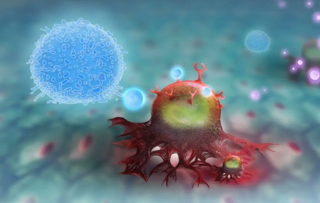
    • Application: This property is used in photodynamic therapy of cancer. , delivered to the tumor and irradiated, causing the death of cancer cells.

    3. Direct impact on pathologies

    • Alzheimer’s Disease: MS is able to inhibit the formation and aggregation of beta-amyloid plaques — one of the main markers of this disease.
    • DNA Repair: Research by Gureev’s group has shown that MS and its metabolite Azur B can trigger signaling pathways responsible for DNA repair, particularly in mitochondria. This explains its protective role in chemotherapy.

    4. Impact on the microbiome

    In high doses, MS exhibits antibacterial properties, which can disrupt the composition of the intestinal microbiome. Modern research necessarily takes into account the gut-brain axis, since changes in the microbiota directly affect cognitive functions.

    Dosages: From therapy to toxicity

    The dose determines whether the MS will be a drug or a poison. The data are primarily based on preclinical animal studies.

    • Low doses (1-4 mg / kg): They were considered effective in early studies. However, current research shows that these doses may not be sufficient for a pronounced therapeutic effect in mouse models.
    • Therapeutic dose (~15 mg / kg): It was at this dosage that the older mice showed significant improvements in memory and cognitive function in the Morris test. This is the dose at which the ‘bypass’ mechanism works effectively, and no serious side effects are observed.
    • Toxic doses (≥50 mg / kg): At such high doses, negative effects begin to appear:
      • Inhibition of cognitive functions.
      • Violation of the composition of the intestinal microbiome.
      • General toxic effects.

    For a person weighing 90 kg, the dose of 15 mg / kg is 1350 mg.
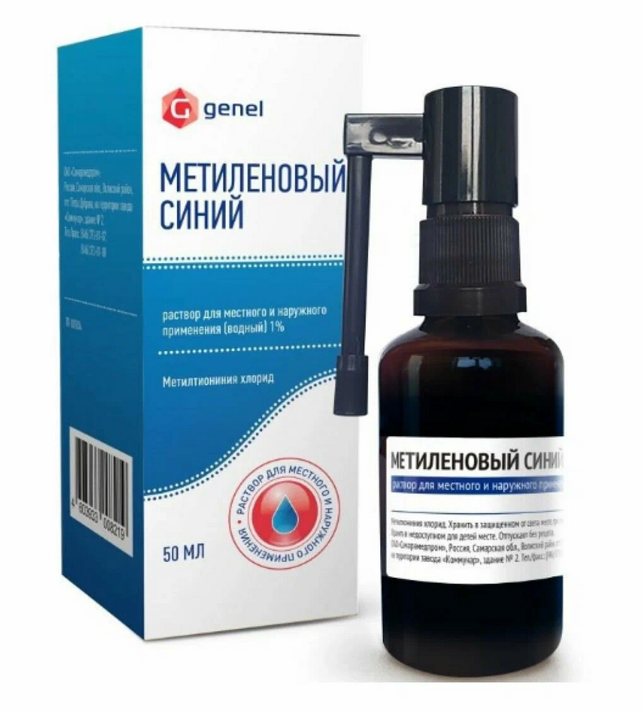
    This is equal to 135 ml of 1% solution, which is approximately 2700 drops.
    This is a HUGE and POTENTIALLY DANGEROUS dose.
    Toxicity: As indicated in the interview with the researcher, in mice, toxic effects (memory suppression, violation of the microbiome) began with a dose of 50 mg/kg. For a 90 kg person, this would be 4,500 mg. Our calculated dose of 1,350 mg (15 mg / kg) is in a potentially risky area, especially without medical supervision.
    Liquid volume: 135 ml is more than half of a standard glass. Drinking such a volume of concentrated dye, which is also a powerful medicine, is an extreme and dangerous procedure for health.
    Conclusion: The 15 mg/kg dose studied in mice is NOT INTENDED for self-administration by humans. It is used in strict preclinical research settings. Self-medication at these dosages can lead to serious side effects, including gastrointestinal damage, dysbiosis, and neurotoxicity.

    A wide therapeutic window (the difference between the effective and toxic dose) makes MS a promising drug.

    Important: Transferring doses from animals to humans is a complex process. These doses are indicative and are given for scientific and informational purposes.

    Side effects and precautions

    • Cosmetic: The most harmless, but noticeable — staining of urine in blue-green color. In rare cases, with prolonged use, a bluish tinge of the sclera of the eyes can be observed.
    • Serious issues: Risk of light toxicity. Taking high doses without understanding the mechanisms can lead to unpredictable consequences.

    Can I take methylene blue for prevention?

    The researcher’s answer is a categorical ‘ no ‘ to self-medication.

    Methylene blue is a powerful medicine, not a dietary supplement.

    Preventive use of MS by healthy people is an unjustified risk. Its use should be strictly within the framework of clinical protocols under the supervision of a doctor after approval by regulatory authorities (for example, the FDA).

    Prospects and conclusions

    Methylene blue is a molecule with huge potential. Today, research is going in several directions:

    1. Neurodegenerative diseases: Attempts to conduct correct clinical trials for Alzheimer’s disease continue.
    2. Synergy with chemotherapy: Its ability to protect healthy tissues (kidneys, brain) from the toxicity of drugs like cisplatin is being studied.
    3. New analogs: Scientists are investigating MS derivatives (Uredin blue), which can be even more effective and safe.

    Conclusion: Methylene blue is not a ‘mind pill’ or an elixir of youth. This is a powerful tool that deeply interferes with cellular energy. Like any powerful tool, it requires respectful and professional handling. The future of MS lies in evidence-based medicine, not in amateur activities in the style of ‘biohacking’.

  • D-димер (фибринолитическая активность) (en translation)

    D-димер (фибринолитическая активность) (en translation)

    Зачем он нужен?

    D-димер – это продукт распада фибрина, который образуется при растворении тромбов (фибринолизе). Анализ используют для:
    Диагностики тромбозов (ТЭЛА, тромбоз глубоких вен).
    Исключения тромбообразования (при низком уровне).
    Контроля лечения антикоагулянтами.
    Оценки риска ДВС-синдрома (диссеминированного свёртывания крови).


    Роль D-димера в организме

    1. Маркер тромбообразования:
    • При формировании тромба активируется фибринолиз – процесс разрушения фибрина.
    • D-димер – это «осколок» распавшегося фибрина, его уровень повышается при наличии тромба.
    1. Отсутствует в норме:
    • В здоровом организме D-димер либо не обнаруживается, либо присутствует в минимальных количествах.

    Симптомы при отклонениях

    При повышенном D-димер (гиперкоагуляция)

    • Тромбоз глубоких вен (ТГВ):
    • Отек, боль и покраснение ноги.
    • Тромбоэмболия лёгочной артерии (ТЭЛА):
    • Одышка, боль в груди, кровохарканье.
    • ДВС-синдром:
    • Кровоточивость, множественные тромбы.
    • Беременность, онкология, COVID-19:
    • Умеренное повышение без явных симптомов.

    При пониженном D-димере

    • Нормальное состояние (исключает активный тромбоз).
    • Дефицит фибринолиза (редко, при генетических нарушениях).

    Нормы D-димера в анализах

    Измеряется в плазме крови (цитрат натрия). Референсы зависят от метода:

    МетодНормаПатология
    ИФА (мкг/мл)< 0.5> 0.5 – риск тромбоза
    Количественный (нг/мл)< 250–500> 500 – требует дообследования

    Примечания:

    • У беременных уровень растет (до 1500 нг/мл в III триместре).
    • После операций/травм временно повышен.
    • У пожилых референсы выше (до 800 нг/мл).

    Когда назначают анализ?

    1. Подозрение на ТЭЛА/ТГВ (в сочетании с УЗИ, КТ).
    2. Контроль ДВС-синдрома.
    3. Мониторинг антикоагулянтной терапии (гепарин, варфарин).

    Что делать при повышенном D-димере?

    1. Исключить тромбоз:
    • УЗИ вен, КТ-ангиография лёгких.
    1. Антикоагулянтная терапия:
    • Гепарин (нефракционированный или низкомолекулярный).
    • Прямые антикоагулянты (апиксабан, ривароксабан).
    1. При ДВС-синдроме:
    • Переливание плазмы, гепарин.

    Примеры интерпретации

    • D-димер = 1200 нг/мл + одышка → срочная КТ для исключения ТЭЛА.
    • D-димер = 300 нг/мл → тромбоз маловероятен.

    Вывод

    D-димер – ключевой маркер фибринолиза и тромбообразования.
    Норма: <500 нг/мл (метод зависимый).
    Повышение:

    • >500 нг/мл → искать тромбоз/ДВС.
    • У беременных/пожилых референсы выше.
      Низкий уровень исключает активный тромбоз.

    Пример: При D-димере 50 нг/мл тромбоз маловероятен, при 1500 нг/мл – нужна срочная диагностика.

  • TNF-альфа (en translation)

    TNF-альфа (en translation)

    Роль TNF-α (фактора некроза опухоли-альфа) в организме

    TNF-α – это провоспалительный цитокин, который играет ключевую роль в:

    Иммунной защите – активирует макрофаги и нейтрофилы для борьбы с инфекциями.
    Апоптозе – запускает гибель поврежденных или раковых клеток.
    Воспалении – усиливает выработку других цитокинов (IL-1, IL-6).
    Метаболизме – влияет на инсулинорезистентность и липолиз.
    Терморегуляции – вызывает лихорадку при инфекциях.


    Симптомы и последствия дисбаланса TNF-α

    При избытке (гипервоспаление)

    • Острые состояния:
    • Сепсис, цитокиновый шторм (шок, полиорганная недостаточность).
    • Тяжелые инфекции (COVID-19, туберкулез).
    • Хронические болезни:
    • Аутоиммунные заболевания (ревматоидный артрит, болезнь Крона).
    • Атеросклероз, диабет 2 типа.
    • Депрессия, нейродегенерация (болезнь Альцгеймера).

    При дефиците (крайне редко)

    • Склонность к инфекциям (особенно внутриклеточным, например, туберкулезу).
    • Нарушение заживления ран.
    • Снижение противоопухолевого иммунитета.

    Примечание: Полное отсутствие TNF-α встречается при редких генетических мутациях.


    Нормы TNF-α в анализах

    Уровень TNF-α измеряется в сыворотке крови методом ИФА. Референсы зависят от лаборатории, но обычно:

    СостояниеУровень TNF-α (пг/мл)
    Здоровые люди< 8.1
    Легкое воспаление8.1–15
    Острое воспаление15–50
    Аутоиммунные болезни/сепсис> 50 (до 1000+)

    Когда назначают анализ?

    • Подозрение на сепсис, аутоиммунные заболевания.
    • Контроль терапии биологическими препаратами (ингибиторами TNF-α).
    • Оценка тяжести COVID-19, ревматоидного артрита.

    Что делать при отклонениях?

    При высоком TNF-α

    1. Медикаментозная терапия:
    • Ингибиторы TNF-α (адалимумаб, инфликсимаб – при ревматоидном артрите).
    • Кортикостероиды (преднизолон).
    1. Натуральные методы:
    • Куркумин, омега-3 (снижают воспаление).
    • Низкоуглеводная диета (уменьшает инсулинорезистентность).

    При низком TNF-α

    • Лечение основного иммунодефицита.
    • Вакцинация (по показаниям).

    Интересные факты

    • TNF-α и рак: в низких дозах защищает от опухолей, в высоких – может стимулировать их рост.
    • Депрессия: хронически повышенный TNF-α нарушает выработку серотонина.

    Вывод

    TNF-α – «страж» иммунитета, но при избытке разрушает ткани.
    Норма: < 8.1 пг/мл (кровь).
    Повышение:

    • >15 пг/мл – острое воспаление,
    • >50 пг/мл – риск полиорганных осложнений.
      Коррекция: биопрепараты, противовоспалительная диета.

    Пример: При ревматоидном артрите уровень TNF-α >20 пг/мл – показание для назначения адалимумаба.