Блог

  • Coenzyme Q10 (CoQ10)

    Coenzyme Q10 (CoQ10)

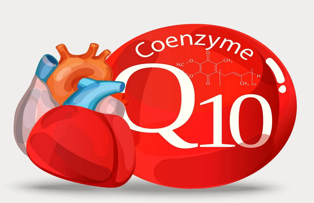
    Coenzyme Q10 (Ubiquinone) — it is a vitamin-like substance that plays a key role in the production of energy (ATP) in the mitochondria and the protection of cells from oxidative stress.

    Main functions:

    1. Energy metabolism -participates in the respiratory chain of mitochondria, helping to synthesize ATP (especially important for the heart, brain, and muscles).
    2. Antioxidant protection -neutralizes free radicals, prevents cell damage.
    3. Heart support -improves myocardial contractility, reduces the risk of atherosclerosis.
    4. Slowing down aging – CoQ10 levels decrease with age.
    5. Immune system -increases the activity of phagocytes.

    Symptoms of CoQ10 deficiency

    A deficit occurs when:

    • Over the age of 40 (natural decrease in synthesis).
    • Taking statins (cholesterol-lowering drugs block CoQ10 synthesis).
    • Chronic diseases (heart failure, diabetes, Parkinson’s disease).

    Signs of a shortage:
    Chronic fatigue, weakness.
    Muscle achesand cramps (especially when taking statins).
    Shortnessof breath, arrhythmia (due to decreased energy in the heart muscle).
    Memory loss, cognitive impairment.
    High blood pressure.


    Symptoms of excess CoQ10

    Overdose is extremely rare, as CoQ10 is non-toxic. Possible effects when taking large doses (>300 mg / day):

    • Nausea, diarrhea.
    • Headache, insomnia.
    • Decreased appetite.

    Note: A natural excess is not possible – the excess is excreted in the bile.


    CoQ10 standards in analyses

    CoQ10 levels are measured in blood plasma or lymphocytes (a more accurate method).

    ParameterStandardOptimal level
    Blood plasma0.5-1.5 mcg / ml>1.0 mcg / ml
    Lymphocytes30-150 nmol / mg of protein>50 nmol / mg

    When is the test scheduled?

    • With muscle weakness while taking statins.
    • For heart failure, migraines.
    • To assess the antioxidant status.

    How to increase CoQ10?

    1. Food:
    • Fatty fish (herring, sardines).
    • Beef, chicken heart.
    • Nuts, seeds, spinach.
    1. Supplements (as directed by your doctor):
    • Ubiquinone (the usual form) – 100-200 mg / day.
    • Ubiquinol (reduced form– — better absorbed, especially after 40 years.
    1. Reducing risk factors:
    • Quitting smoking (accelerates oxidative stress).
    • Blood sugar control (diabetes lowers CoQ10).

    Conclusion

    CoQ10 is a vital component for energy and antioxidant protection.
    Deficiency is manifested by fatigue, muscle weakness, and heart problems.
    , Blood norm: 0.5-1.5 mcg / ml (plasma), > 50 nmol / mg (lymphocytes).
    Supplements are effective for proven deficiencies or statin use.

    Example: If the CoQ10 level is < 0.5 mcg / ml, it is recommended to take 100-200 mg/day in the form of ubiquinol.

  • Antibodies to tissue transglutaminase (anti-tTG)

    Antibodies to tissue transglutaminase (anti-tTG)

    Why is it needed?

    This test is used to diagnose celiac disease– an autoimmune disease in which eating gluten (wheat, rye, and barley protein) causes damage to the lining of the small intestine.

    Main objectives of the analysis:
    ✔ Confirmation of the diagnosis of celiacdisease.
    ✔ Monitoring compliance with a gluten-free diet.
    ✔ Screening for people with a family history of celiacdisease.


    What is Tissue transglutaminase (tTG)?

    • An enzymeinvolved in tissue repair.
    • In celiac disease, the immune system mistakenly attacks tTG, mistaking it for a threat (due to a reaction with gluten).
    • In response , antibodies (IgA and IgG)are produced, which are detected in the analysis.

    Symptoms of a positive test (celiac disease)

    Classic symptoms (in children and adults):

    • Chronic diarrhea or constipation.
    • Bloating, abdominal pain.
    • Weight loss (despite a normal diet).
    • Steatorrhea (greasy, fetid stools).

    Extra-intestinal manifestations:

    • Anemia (due to iron and folic acid deficiency).
    • Osteoporosis (calcium malabsorption).
    • Rash (dermatitis herpetiformis).
    • Infertility, neurological disorders.

    Is there a shortage or excess of tTG?

    • Transglutaminase deficiency -does not occur, as it is an important enzyme of the body.
    • Excess tTG – has no clinical significance, only the presence of antibodies to itis important.

    TTG Antibody standards

    The test is performed in the blood (more often IgA antibodies are determined, less often IgG).

    Type of antibodyNorm (U / ml)Interpretation
    IgA anti-tTG< 15Negative (normal)
    15–30Weakly positive (repeated test)
    > 30Positive (celiac disease is likely)
    IgG anti-tTG (for IgA deficiency)< 15Standard
    > 15Suspected celiac disease

    Important:

    • 5-10% of people with celiac disease may be deficient in IgA, so IgG is additionally tested.
    • Gluten should not be excluded before the analysis – this may give a false negative result.

    What should I do if I get a positive result?

    1. Consultation with a gastroenterologist.
    2. Endoscopy with biopsy (gold standard of diagnostics).
    3. Complete elimination of gluten (wheat, rye, barley).
    4. Control of antibody levels after 6-12 months (to assess the effectiveness of the diet).

    When is the test scheduled?

    • For symptoms of malabsorption (diarrhea, weight loss).
    • If relatives have celiac disease.
    • For autoimmune diseases (type 1 diabetes, autoimmune thyroiditis).
    • For unexplained anemia or osteoporosis.

    Conclusion

    TTG antibodies are a key marker of celiac disease.
    , The norm: < 15 U / ml (IgA).
    A positive result (>30 U/ml) requires a bowel biopsy.
    , Treatment: a strict gluten-free diet for life.

  • Epigenetic age (DNA methylation test)

    Epigenetic age (DNA methylation test)

    Epigenetic age (DNA methylation test): why is it needed?

    Epigenetic age is the biological age of cells, determined by the patterns of DNA methylation (chemical labels on genes that regulate their activity). Unlike chronological age, it reflects the actual rate of aging of the body and the risks of age-related diseases.

    How does DNA methylation work?

    • With age, some genes are ‘turned off ‘(hypermethylation), while others are ‘turned on’ (hypomethylation).
    • These changes affect:
    • Inflammation (IL-6, TNF-α).
    • DNA repair (p16, p53).
    • Mitochondrial function (PPARGC1A).
    • Accelerated methylation = rapid aging.

    Why do I need this test?

    1. Estimation of the aging rate:
    • If the epigenetic age is higher than the chronological age– the risk of diabetes, cancer, or dementia is increased.
    • If lower , it indicates a ‘young’ biological age.
    1. Personalized prevention:
    • Lifestyle correction (diet, sports, sleep).
    • Reception of methyl donors (folate, B12) with impaired methylation.
    1. Prognosis of therapy effectiveness:
    • For example, people with ‘young’ epivozrastom better take root transplants.

    Symptoms of accelerated epigenetic aging

    There are no direct symptoms, but indirect signs:

    • Early wrinkles, gray hair.
    • Chronic fatigue, cognitive decline.
    • Common inflammatory diseases (arthritis, atherosclerosis).
    • Insulin resistance (prediabetes).

    Risk factors for accelerated aging:

    • Smoking, alcohol.
    • Obesity, stress.
    • Vitamin B9/B12 deficiency.

    Symptoms of ‘delayed’ aging

    • High physical and mental activity in the elderly.
    • No chronic diseases (for example, normal blood pressure at age 70).
    • Fast recovery after loads.

    Norms in analysis

    Epigenetic age is measured in years and compared with chronological age:

    • Optimal: biological age < chronological age.
    • Accelerated aging: +3-5 years or more.
    • Delayed aging: -3-5 years or more.

    Sample result:


    Different ‘ clocks ‘(Horvath, Hannum, PhenoAge) evaluate different aspects of aging.


    What to do with accelerated aging?

    1. Diet correction:
    • Methyl donors: leafy greens, liver, and eggs (sources of folate and B12).
    • Antioxidants: berries, curcumin, omega-3.
    • Limit sugar and trans fats.
    1. Lifestyle:
    • Intermittent fasting (activates autophagy).
    • Physical activity (strength + cardio).
    • Sleep 7-9 hours (DNA repair).
    1. Supplements (as directed by your doctor):
    • NMN/NR (precursors of NAD+).
    • Resveratrol (sirtuin activator).
    • Methylfolate (with impaired methylation).

    Where is the test used?

    • Anti-age medicine -selection of anti-aging programs.
    • Oncology -assessment of the aggressiveness of tumors (for example, gliomas).
    • Longevity research – study of the effects of cryonics, fasting.

    Conclusion

    • Epigenetic age is an accurate marker of biological aging based on DNA methylation.
    • The norm: matching or lagging behind the chronological age.
    • Accelerated aging is corrected by diet, exercise, and methyl donors.
    • The test is useful for early prevention of age-related diseases and a personalized approach to health.
  • Calprotectin

    Calprotectin

    Analysis of feces for calprotectin

    Why is it needed?

    Calprotectin is a proteinthat is released by white blood cells (neutrophils) during inflammation in the intestines. Its level in the stool helps distinguish functional disorders (such as IBS) from inflammatory bowel diseases (IBD):
    Crohn‘s disease.
    Ulcerative colitis.
    Intestinal infections.
    Diverticulitis, intestinal tumors.


    What does calprotectin do in the body?

    • Inflammatory marker: increases when immune cells in the intestinal mucosa are activated.
    • Protective function: binds zinc and calcium, limiting bacterial growth.
    • It does not depend on diet or medication (except for NSAIDs, which may increase its level).

    Symptoms with increased calprotectin

    An excess indicates active inflammation in the intestines:

    • Chronic diarrhea or constipation.
    • Abdominal pain, cramps.
    • Blood or mucus in the stool.
    • Weight loss, weakness.

    Reasons for the increase:

    • IBD (Crohn’s disease, ulcerative colitis).
    • Intestinal infections (salmonellosis, campylobacteriosis).
    • Celiac disease in the active phase.
    • Taking NSAIDs (ibuprofen, aspirin).

    Is there a calprotectin deficiency?

    No, a low level is the norm.

    • If calprotectin is normal, but there are symptoms – probably a functional disorder (IBS).

    Calprotectin norms in feces

    Level (mcg/g)Interpretation
    <50Normal (no inflammation)
    50–120Moderate elevation (requires monitoring)
    >120High inflammation (suspected IBD)

    Notes:

    • In children under 4 years of age, the norm is higher (up to 150 mcg/g).
    • At the level of 50-120 mcg/g , the analysis is repeated after 1-2 months.

    What to do with increased calprotectin?

    1. Consultation with a gastroenterologist.
    2. Additional surveys:
    • Colonoscopy with biopsy.
    • Analysis for infections (fecal culture, PCR).
    1. Treatment of the underlying disease:
    • In IBD – anti-inflammatory drugs (mesalazine, biological therapy).
    • In case of infection – antibiotics.

    When is the test scheduled?

    • Suspected IBD (with chronic diarrhea, pain).
    • Monitoring treatment for Crohn’s disease or ulcerative colitis.
    • Differential diagnosis of IBS and IBD.

    Conclusion

    Calprotectin is a reliable marker of intestinal inflammation.
    , The norm: < 50 mcg / g** (for children under 4 years – up to * * 150 mcg/g**). **The increase (>120 mcg/g) requires an in-depth examination.
    A low level rules out serious inflammation (most likely IBS).

    Example: If the result is 200 mcg / g, a colonoscopy is needed to check for Crohn’s disease.

  • Feces for elastase-1

    Feces for elastase-1

    Stool analysis for elastase-1

    Why is it needed?

    Elastase-1 is an enzyme of the pancreasthat is involved in the digestion of proteins. Its level in the stool reflects the exocrine function of the pancreas (the ability to produce digestive enzymes).

    The analysis is prescribed for the diagnosis of:
    Chronic pancreatitis (decreased production of enzymes).
    Cystic fibrosis (a genetic disorder involving the pancreas).
    Malabsorption syndrome (malabsorption of nutrients).
    Bowel diseases (celiac disease, Crohn’s disease).


    Symptoms of Elastase-1 deficiency

    If there is a lack of the enzyme, the pancreas can not cope with the digestion of food, which leads to:

    • Greasy, fetid stools (steatorrhea).
    • Bloating, gas.
    • Weight loss (despite a normal diet).
    • Lack of vitamins A, D, E, and K (due to poor fat absorption).
    • Chronic diarrhea.

    Causes of low Elastase-1:

    • Chronic pancreatitis.
    • Cystic fibrosis.
    • Age-related decline in pancreatic function.
    • Pancreatic surgery.

    Can there be an excess of elastase-1?

    No, elevated elastase-1 levels are not clinically relevant.

    • If the result is higher than normal, this does not indicate a disease.
    • Sometimes this happens with diarrhea of non-pancreatic origin (for example, with infections).

    Norm of elastase-1 in feces

    Level (mcg/g of feces)Interpretation
    >500Norm (optimal level)
    200–500Mild / moderate insufficiency
    <100Severe pancreatic insufficiency

    Important:

    • The test is given without preparation (it does not require a diet or medication withdrawal).
    • Feces are collected in a sterile container (1-2 g is enough).
    • Do not freeze the sample!

    What to do with low elastase-1?

    1. Enzyme replacement therapy (Creon, Pancreatin).
    2. A low-fat diet (to reduce the burden on the pancreas).
    3. Vitamin control (taking fat-soluble vitamins A, D, E, and K).
    4. Additional surveys:
    • Ultrasound/MRI of the pancreas.
    • Test for cystic fibrosis (if the cause is unknown).

    When is the test scheduled?

    • Chronic diarrhea, steatorrhea.
    • Unexplained weight loss.
    • Suspected pancreatitis or cystic fibrosis.

    Conclusion

    Fecal elastase-1 is a reliable marker of pancreatic function.
    , The norm: > 500 mcg/g
    . Дефицит Deficiency (<200 mcg/g) indicates pancreatic insufficiency.
    Treatment: Enzymes + diet + vitamin control.

  • Detoxification Genetic Polymorphism Test

    Detoxification Genetic Polymorphism Test

    This test analyzes the genes responsible for the work of detoxification enzymes (phase I and II), which neutralize toxins, drugs, carcinogens and metabolic products. It helps to identify congenital disorders that increase the risk of chronic diseases, intoxication, and drug intolerance.

    What genes are being tested?

    1. Phase I (Cytochrome P450):
    • CYP1A1, CYP1B1 – metabolism of carcinogens (for example, benzopyrene from tobacco smoke).
    • CYP2D6 -neutralization of 25% of drugs (antidepressants, beta-blockers).
    • CYP2C9, CYP2C19 -metabolism of warfarin, NSAIDs, and antidepressants.
    1. Phase II (conjugation):
    • GSTM1, GSTT1 – deactivation of toxins and free radicals (if fission occurs, there is a risk of cancer).
    • COMT – destruction of catecholamines (epinephrine, dopamine).
    • NAT2 -neutralization of aromatic amines (in case of coffee and tea intolerance).

    Why do I need this test?

    1. Risk assessment:
    • Oncology (with weak detoxification of carcinogens).
    • Chronic intoxication (heavy metals, pesticides).
    • Drug intolerance (for example, slow metabolism of CYP2D6 → codeine poisoning).
    • Hormonal disorders (COMT affects estrogens and dopamine).
    1. Personalized recommendations:
    • Selection of safe medications and doses.
    • Diet correction (for example, for NAT2 polymorphisms, smoked products are excluded).
    • Appointment of antioxidants (for GST defects).

    Symptoms of detoxification disorders

    With weak enzyme activity (accumulation of toxins):

    • Chronic fatigue, headaches.
    • Allergies, skin rashes (eczema, acne).
    • Intolerance to alcohol, coffee, and medications.
    • Increased risk of cancer (especially lung, breast, and bladder).
    • Hormonal failures (PMS, endometriosis — with slow COMT).

    With hyperactivity of enzymes (rapid breakdown of useful substances):

    • Low effectiveness of painkillers or antidepressants.
    • Vitamin D deficiency (due to accelerated CYP2R1 metabolism).
    • Lack of dopamine (with rapid COMT — > anxiety, ADHD).

    Norms in analysis

    A genetic test has no ‘norms’ in the traditional sense. The result shows the type of polymorphism:

    • Fast metabolism (high enzyme activity).
    • Slow metabolism (risk of accumulation of toxins).
    • Normal type (optimal function).

    Sample result:


    What should I do when detecting polymorphisms?

    1. Avoid toxins:
    • Abstaining from smoking and alcohol.
    • Restriction of smoked products (at NAT2).
    • Use of eco-friendly cosmetics.
    1. Detoxification Support:
    • Glutathione (for GST defects).
    • Magnesium, B12, folate (for the work of cytochromes).
    • Indole-3-carbinol (for slow COMT and high estrogens).
    1. Personal selection of medicines:
    • For’ slow metabolizers ‘ – lower doses.
    • For’ fast ‘ – alternative drugs.

    Examples of the impact of polymorphisms

    The genePolymorphismEffectsRecommendations
    CYP2C192/2Ineffectiveness of clopidogrelReplacement with ticagrelor
    COMTVal158MetAnxiety, low dopamineSAM-e, magnesium
    GSTM1DeletionLung cancer riskCurcumin, selenium

    Conclusion

    • The detoxification polymorphism test identifies individual risks of diseases and drug reactions.
    • Symptoms of violations: chronic intoxication, drug intolerance, hormonal disorders.
    • ‘Norms’ is the definition of the type of metabolism (fast/slow).
    • Correction: diet, antioxidants, personalized pharmacotherapy.

    The test is especially useful when:

    • Unexplained chronic symptoms.
    • Family history of oncology.
    • Frequent side effects from medications.
  • Progesterone and estradiol (in women)

    Progesterone and estradiol (in women)

    The role of progesterone and estradiol in women

    1. Estradiol (E2)

    Estradiol is the main estrogen produced in the ovaries (follicles), as well as in the adrenal glands and adipose tissue.

    Functions:
    Regulation of the menstrual cycle -stimulates the growth of the endometrium.
    Development of secondary sexual characteristics (breast, female body type).
    Strengthen bones (prevents osteoporosis).
    It affects the skin and hair (increases collagen, hydration).
    Pregnancy Support -prepares the uterus for implantation.


    2. Progesterone

    Progesterone is a hormone of the yellow body of the ovaries and placenta (during pregnancy).

    Functions:
    Preparation of the endometrium for implantation of a fertilized egg.
    Reduced uterine tone (prevents miscarriage).
    Suppressing ovulation in the second phase of the cycle and during pregnancy.
    Effect on the mammary glands (preparation for lactation).


    Norms in analyses

    Estradiol (E2)

    Cycle phaseNorm (pg / ml)Norm (pmol / l)
    Follicular (1-14 days)20–15070–550
    Ovulation (~day 14)100–400370–1470
    Luteal acid(15-28 days)50–220180–800
    Menopause<20<70

    Progesterone

    Cycle phaseNorm (ng / ml)Norm (nmol / l)
    Follicular0.1–0.80.3–2.5
    Ovulation0.1–1.50.3–4.8
    Luteal acid2–256–80
    Menopause<0.1<0.3
    Pregnancy (I trimester)10–4430–140

    Symptoms of deficiency and excess

    Estradiol

    Deficiency (hypoestrogenism)Excess (hyperestrogenism)
    влагали Vaginal dryness, painful sex✅ Breast swelling and swelling
    Hot flashes, sweating (menopause)✅ Increased anxiety
    ✅ Brittle bones (osteoporosis)✅ Migraines, PMS
    ✅ Dry skin, wrinkles Fibrocystic mastopathy
    цикла Cycle disorders (amenorrhea)✅ Risk of endometriosis, fibroids

    Causes of the deficit:

    • Menopause, early menopause.
    • Ovarian depletion (PCOS, stress).
    • Strict diets (anorexia).

    Causes of excess:

    • Obesity (adipose tissue produces estrogens).
    • Hormone-producing tumors.
    • Taking estrogen-containing medications.

    Progesterone

    DeficitExcess
    ✅ Irregular monthly periods✅ Drowsiness, fatigue
    Infertility, miscarriages✅ Vertigo
    ✅ PMS (irritability)✅ Decreased libido
    ✅ Dry skin, hair loss✅ Hypotension (low blood pressure)

    Causes of the deficit:

    • Insufficiency of the yellow body.
    • Chronic stress (cortisol ‘steals’ progesterone).
    • Hypothyroidism.

    Causes of excess:

    • Pregnancy.
    • Yellow body cysts.
    • Taking progesterone medications (Duphaston, Utrozhestan).

    When should I get tested?

    • Estradiol3-5 day of the cycle (follicular phase) or in dynamics.
    • Progesterone21-23 days (with a 28-day cycle) in the luteal phase.

    Additional tests:

    • FSH, LH – to assess ovulation.
    • Testosterone, DHT -for hirsutism.
    • TSH – if hypothyroidism is suspected.

    How do I normalize my levels?

    With estradiol deficiency:

    • Phytoestrogens (soy, flax, red clover).
    • HRT (estrogen-containing drugs).
    • Stress reduction (cortisol suppresses estrogens).

    If there is an excess of estradiol:

    • Fiber (removes excess).
    • Indole-3-carbinol (broccoli, cabbage).

    With progesterone deficiency:

    • Vitamin B6, magnesium (yellow body support).
    • Progesterone cream / tablets.

    With an excess of progesterone:

    • It usually does not require correction (except in cases of cysts or medication).

    Conclusion

    Estradiol — the hormone of femininity, affects the cycle, skin, bones.
    Progesterone is a pregnancy hormone that protects against miscarriages.
    The norms depend on the phase of the cycle (especially the luteal peak of progesterone is important).
    An imbalance leads to PMS, infertility, and early aging.
    Correction – diet, HRT, stress reduction.

  • ACTG

    ACTG

    Adrenocorticotropic hormone (ACTH): role, symptoms of blood imbalance and normality

    1. The role of ACTH in the body

    ACTH (adrenocorticotropic hormone) is a hormone produced by the anterior pituitary gland under the control of the hypothalamus (via corticoliberin, CRH).

    Main functions:
    Adrenal Stimulation – ACTH causes the adrenal cortex to produce:

    • Cortisol (a glucocorticoid that regulates metabolism and stress response).
    • Androgens (precursors of sex hormones).
    • To a lesser extent – aldosterone (a mineralocorticoid that controls the water-salt balance).

    Maintaining circadian rhythms – ACTH levels are maximal in the morning (6-8 hours) and minimal in the evening.

    Participation in the stress response – under stress (physical or emotional), ACTH secretion increases dramatically.


    2. Symptoms of ACTH deficiency (hypocorticism)

    Reasons:

    • Hypopituitarism (pituitary insufficiency due to a tumor, injury, or ischemia).
    • Long-term use of glucocorticoids (suppresses the production of ACTH on the feedback principle).
    • Sheehan’s syndrome (postpartum pituitary necrosis).

    Symptoms:

    • Chronic weakness, fatigue (due to lack of cortisol).
    • Hypoglycemia (low blood sugar).
    • Weight loss, lack of appetite.
    • Hypotension (low blood pressure).
    • Pallor of the skin (due to a decrease in ACTH and melanocyte-stimulating hormone).

    3. Symptoms of excess ACTH (hypercorticism)

    Reasons:

    • Itsenko-Cushing ‘s disease(pituitary adenoma, excessively producing ACTH).
    • Ectopic production of ACTH (in small cell lung cancer, carcinoids).
    • Adrenal insufficiency (the adrenal glands do not respond to ACTH — > compensatory growth of ACTH).

    Symptoms:

    • Obesity with fat deposition on the face (‘moon face’), stomach, neck.
    • Stretch marks (striae) purple color on the skin.
    • Arterial hypertension (high blood pressure).
    • Osteoporosis (brittle bones).
    • Hirsutism and acne (due to excess androgens).
    • Impaired glucose tolerance (prediabetes).

    4. Norms of ACTH in the blood test

    ACTH levels fluctuate strongly throughout the day:

    Time to submit the analysisReference values (pg / ml)
    Morning (7: 00-10: 00)7-63 (maximum level)
    Evening (18: 00-22: 00)< 30 (minimum level)

    Important:

    • The test is taken strictly on an empty stomach, in the morning, at rest (stress distorts the result).
    • At the same time, cortisol is checked for a comprehensive assessment.

    5. When is an ACTH test scheduled?

    • Suspected Itsenko-Cushing’s disease / syndrome.
    • Diagnosis of adrenal insufficiency (Addison’s disease).
    • Evaluation of the hypothalamic-pituitary-adrenal axis.
    • Control of long-term glucocorticoid therapy.

    In case of abnormalities , an endocrinologist’s consultation and additional tests (corticoliberin test, pituitary MRI) are required.

  • Genetic test for folate metabolism

    Genetic test for folate metabolism

    This test analyzes genes that affect the absorption of folic acid (vitamin B9) and its conversion to the active form (5-MTHF). It helps to identify congenital disorders that prevent the body from properly using folate, even with sufficient intake from food.

    What genes are being tested?

    1. MTHFR (C677T and A1298C) — most important:
    • Mutation C677T reduces the activity of the enzyme by 70%, which leads to the accumulation of homocysteine.
    • A1298C causes less severe disorders.
    1. MTR and MTRR -affect the remethylation of homocysteine.
    2. DHFR -responsible for folic acid recovery.

    Why do I need this test?

    1. Risk assessment:
    • Thrombosis (due to increased homocysteine).
    • Pregnancy complications (miscarriages, neural tube defects in the fetus).
    • Depression, dementia (impaired neurotransmitter synthesis).
    • Anemia (megaloblastic due to hematopoietic defects).
    1. Personalization of treatment:
    • Selection of the folate form (regular folic acid may not be absorbed with MTHFR mutations).
    • Dosage of vitamins B12, B6 and B9 to reduce homocysteine.

    Symptoms of folate deficiency

    They occur due to a violation of DNA synthesis and an excess of homocysteine:

    • General information: weakness, pallor, dizziness.
    • Neurological: irritability, depression, cognitive impairment.
    • Hematological: megaloblastic anemia (large immature red blood cells).
    • Pregnancy: malformations of the fetus (spina bifida, anencephaly).
    • Cardiovascular diseases: early atherosclerosis, thrombosis.

    Risk groups:

    • People with a family history of thrombosis or miscarriage.
    • Vegans (B12 deficiency worsens folate deficiency).
    • Patients with autoimmune diseases (celiac disease, Crohn’s disease).

    Symptoms of excess folate

    Rare, but possible with an overdose of synthetic folic acid:

    • Masking B12 deficiency (folic acid ‘corrects’ anemia, but does not stop nerve damage).
    • Allergic reactions (skin rash, itching).
    • Increased excitability of the central nervous system (insomnia, irritability).
    • Growth of tumors (theoretical risk for pre-existing neoplasms).

    Blood folate levels

    Analysis for folate is performed in serum or red blood cells (a more accurate indicator).

    ParameterStandard
    Serum Folate7-45 nmol / l (3-20 ng / ml)
    Folate in red blood cells360-1400 nmol / l (160-620 ng / ml)
    Homocysteine<15 micromol/l (optimal <8)

    For MTHFR mutations:

    • Folate levels may be normal, but homocysteine levels are elevated (>10-15 mmol / l).
    • Red blood cell folate is a more reliable marker than serum folate.

    What should I do if mutations are detected?

    1. Replace folic acid with methylated forms:
    • 5-MTHF (methylfolate) — is absorbed even with the MTHFR mutation.
    • Dosage: 400-1000 mcg / day (for pregnancy up to 5 mg).
    1. Combine with vitamins B12 and B6:
    • B12 (methylcobalamin) – 500-1000 mcg / day.
    • B6 (pyridoxal-5-phosphate) – 25-50 mg / day.
    1. Control homocysteine:
    • Target level: <7-8 mmol/l.
    1. Diet:
    • Leafy greens (spinach, arugula).
    • Liver, eggs, and legumes.
    • Avocado, citrus fruits.

    Example of a genetic report


    Conclusion

    • A genetic test reveals the risk of folate metabolism disorders leading to increased homocysteine.
    • The deficiency is manifested by anemia, neurological and obstetric complications.
    • Excess is possible only when taking synthetic forms.
    • Folate norms: 7-45 nmol/l in serum, homocysteine <8 mmol/l.
    • Treatment: methylfolate + B12 + B6 under the control of assays.

    The test is especially important for those planning pregnancy and patients with a history of thrombosis.

  • Light chain neurofilament

    Light chain neurofilament

    The role of light chain neurofilament (NfL) in the body

    Neurofilament Light Chain (NfL) is a structural protein of neurons that is part of their cytoskeleton. It plays a key role in maintaining the shape and stability of axons (long processes of nerve cells).

    Key NfL Features:

    1. Maintaining the structure of axons -provides mechanical strength and flexibility of nerve fibers.
    2. Participation in intracellular transport -helps to move substances inside the neuron.
    3. Neural tissue damage marker – when neurons are destroyed, NfL is released into the cerebrospinal fluid and blood.

    When does the NfL become an important metric?

    The level of NfL in the blood or cerebrospinal fluid is used as a biomarker of neurodegenerative and neurological diseases, since its increase correlates with neuronal damage.

    Diseases associated with increased NfL:

    • Multiple sclerosis– MS) is a marker of demyelination activity.
    • Alzheimer ‘s disease and other dementias.
    • Parkinson ‘s disease and atypical parkinsonism.
    • Amyotrophic lateral sclerosis (ALS).
    • Traumatic brain injuries and strokes.
    • HIV-associated neurocognitive dysfunction.

    Symptoms associated with Changing NfL Levels

    NfL is not a cause of disease, but an indicator of neuronal damage. Therefore, ‘symptoms’ refer to the main diseases:

    With increased NfL (neurodegeneration or injury):

    • Cognitive disorders (problems with memory, thinking).
    • Muscle weakness, tremor, poor coordination.
    • Numbness, tingling in the extremities (in MS).
    • Progressive dementia (with Alzheimer’s).

    NfL Deficit

    A physiologically low level of NfL is normal, but a complete lack of protein is impossible, since it is necessary for the structure of neurons.


    NfL norm in a Blood test

    NfL levels are measured in picograms per milliliter (pg / ml). The reference values depend on the age and method of analysis (for example, the Simoa method is the most sensitive).

    Approximate standards:

    AgeBlood NfL level (pg / ml)
    < 40 years old<10
    40-60 years old<15
    > 60 years old<20–30

    Important:

    • Children have a higher level of NfL than adults (due to the active development of the nervous system).
    • In neurological diseases, the values can exceed 50-100 pg / ml and even reach 1000 pg / ml (in late-stage ALS).

    How is the analysis performed?

    1. Material: blood or CSF (cerebrospinal fluid).
    2. Method: immunoassay using antibodies (for example, Single Molecule Array-Simoa).
    3. When they are assigned:
    • Diagnosis and monitoring of multiple sclerosis.
    • Assessment of the progression of neurodegenerative diseases.
    • Monitoring the effectiveness of therapy.

    What to do with an increased NfL?

    1. Consult a neurologist to find the cause (MRI, lumbar puncture, etc.).
    2. Treatment of the underlying disease (for example, immunotherapy for MS).
    3. Dynamic follow – up-repeated tests to assess the rate of progression.

    Conclusion

    • NfL protein is a marker of neuronal damage, not an independent disease.
    • The norm: depends on the age, on average < 10-30 pg / ml in the blood.
    • An increase indicates neurodegeneration, trauma, or autoimmune processes (MS, ALS, Alzheimer’s).
    • NfL analysis is used in neurology for early diagnosis and disease control.

    If the NfL level is elevated, an in-depth examination by a specialist is required.