Рубрика: Medical articles>General articles

  • Understanding the size parameters of peptides and amino acids

    Understanding the size parameters of peptides and amino acids

    Amino Acid Size

    The average molecular weight (based on the weight of a hydrogen atom equal to 1) of each amino acid is approximately 100 to 125 daltons.

    Peptide Size

    The size of the peptide plays an important role in its ability to penetrate the skin. In general, peptides up to 500 daltons (Da)in size they can penetrate the upper layers of the skin, such as the epidermis.

    However, for the peptide to enter the deeper layers of the dermis or the bloodstream, it must be small enough and sufficiently lipophilic (i.e., fat-soluble).

      • Molecules up to 500 daltons in size often penetrate the top layer of the skin (the stratum corneum of the epidermis), but their penetration into the dermis is limited.
      • Peptides with a molecular weight greater than 1000 daltons (for example, larger peptides or proteins) often do not penetrate deep into the skin and remain in the upper layers of the epidermis, not reaching the dermis.

    Thus, peptides up to 500 Da in size can penetrate the skin and have effects such as moisturizing, stimulating collagen synthesis, and other cosmetic or therapeutic effects. Peptides of a larger mass are likely to remain on the surface or in the upper layers of the epidermis.

    Protein sizes

    Proteins are polymer molecules in which amino acids serve as monomers. In the composition of proteins in the human body, only 20 alpha-amino acids are found. The same amino acids are present in proteins with different structures and functions. The individuality of protein molecules is determined by the sequence of amino acids in the protein.

    Amino acids are organic acids that contain one or more amino groups.

    All alpha-amino acids, except for aminoacetic acid (glycine), have an asymmetric a-carbon atom and exist as two enantiomers.Almost all proteins are made up of 20 α-amino acids belonging to the K-series, with the exception of glycine.

  • Stimulating autophagy

    Stimulating autophagy

    Autophagy stimulation is a key area in longevity and health research. The body has a complex system of regulation of this process, and it can be influenced by both natural and (potentially) pharmacological methods.

    Here are the main mechanisms and factors that stimulate autophagy and improve the functioning of lysosomes.

    1. Energy stress (‘Turning on hunger’)

    This is the most powerful and natural incentive. When a cell doesn’t get enough energy, it turns on a ‘recycling mode’ to get rid of the old one and produce new energy.

    • Potassium starvation: The most effective method. The lack of nutrients (especially amino acids and glucose) leads to a drop in blood levels.:
      • Insulin and IGF-1 (insulin-like growth factor 1).
      • mTOR (target of rapamycin in mammals). mTOR is the main ‘brake’ of autophagy. When its activity is high (with an abundance of food), the cell grows and divides, and autophagy is suppressed. Fasting slows down mTOR, removing this ‘brake’ and triggering autophagy.
      • Activation of AMPK (AMP-activated protein kinase). This is the’ energy level sensor ‘ of the cell. When the level of ATP (the energy currency of the cell) is low, AMPK is activated and involves energy production processes, including autophagy. AMPK directly suppresses mTOR.
    • Interval fasting and diets that mimic fasting: More practiced approaches that give the same effect as prolonged fasting, but in a milder form.

    2. Physical activity

    Physical activity creates moderate stress for muscle cells and other organs.

    • Mechanical damage and oxidative stress: During exercise, damaged proteins and organelles are formed in the cells. This serves as a direct signal for their utilization through autophagy.
    • Energy consumption: Exercise depletes glycogen and ATP stores, which, like fasting, activates AMPK and inhibits mTOR.

    3. Cellular stress

    Any moderate stress can ‘shake up’ the cell and start cleaning processes.

    • Oxidative stress: The formation of reactive oxygen species (ROS) serves as a signal to trigger autophagy.
    • Hypoxia (lack of oxygen): Activates special proteins (HIF) that can induce autophagy.
    • Heat stress: Rising temperatures are also a trigger.

    How to improve the performance and number of lysosomes?

    Stimulating autophagy is only half the battle. It is important that the ‘processing plants’ — lysosomes-work efficiently.

    1. Synthesis of new lysosomes (Lysosomogenesis)

    A cell can increase the number of lysosomes when it realizes that the need for recycling has increased. The main ‘regulator of lysosomes’ — transcription factor EB (TFEB) — is responsible for this process.

    • How is TFEB activated?
      • During starvation and stress, when mTOR is inactive, TFEB moves from the cytoplasm to the nucleus.
      • In the nucleus, it ‘sits’ on genes encoding lysosomal enzymes (cathepsins) and lysosomal membrane proteins themselves.
      • This leads to a massive synthesis of new,’ fresh ‘ lysosomes and enzymes for them.

    Factors that activate TFEB (and hence lysosomogenesis):

    • Same as for autophagy: fasting, exercise, oxidative stress.
    • Some polyphenols, such as curcumin and resveratrol, can also promote TFEB activation.

    2. ‘Rejuvenation’ of existing lysosomes

    For lysosomes to work well, you need to maintain their internal environment.

    • Acidity (pH): Lysosomes require an acidic environment (pH ~4.5-5.0) to activate enzymes. With age and with diseases (for example, neurodegenerative diseases), the pH in lysosomes can increase, and enzymes stop working.
      • What helps? Adequate functioning of the proton pump (V-ATPase), which pumps protons into the lysosome. It requires energy (ATP) and certain lipids to work.
    • Zinc and other cofactors: Some lysosomal enzymes require zinc and other trace elements to function.

    Practical conclusions: what ‘helps’ autophagy and lysosomes?

    StrategyMechanism of action
    Interval fasting (for example, 16/8)Insulin/glucose reduction, mTOR inhibition, AMPK and TFEB activation.
    Regular physical activityEnergy consumption, AMPK activation, direct stress from damage.
    A protein-restricted diet (especially animal-based)Decreased mTOR activity (leucine and other amino acids are powerful mTOR stimulants).
    Consumption of foods rich in polyphenols (green tea, turmeric, berries, red grapes)Antioxidant effect, potential activation of pathways leading to autophagy (sirtuins, TFEB).
    Healthy sleepDuring sleep, brain cleaning processes are activated, including autophagy (glymphatic system).
    Avoiding constant ‘snacking’Constant food intake supports high insulin levels and mTOR activity by suppressing autophagy.

    Important warning: Autophagy is a double-edged sword. Its chronic or excessive activation can be harmful. The ‘more is better’ approach doesn’t work here. Stimulation should be cyclical and moderate (for example, through fasting) to give the cell time to recover.

  • Iron in the body and Hemosiderin

    Iron in the body and Hemosiderin

    In a living organism, there are practically no ‘free’ metal ions in significant quantities, because they are toxic. In the liver (and other organs), iron accumulates NOT in a free form, but in a bound, non-toxic and bioavailable form.

    Why is free iron dangerous?

    Free iron ions (Fe2⁺ and Fe3⁺) in the cell act as a powerful oxidative stress catalyst (pro-oxidizer). They are involved in the Fenton reaction, during which extremely active and dangerous free radicals are formedthat damage the skin.:

    • Cell membrane lipids
    • Squirrels
    • DNA

    This can lead to cell death and organ failure.

    To prevent this from happening, the body has a special system for storing and transporting iron, where it is always associated with special proteins.

    The main forms of iron storage in the liver

    Iron in the liver is stored in two main protein ‘containers’:

    1. Ferritin (Main storage form)

    It is the main and non-toxic protein depot of iron in our body.

    • Structure: Ferritin is similar to a hollow ball (nanometer ‘safe’), consisting of 24 subunits.
    • Capacity: Up to 4,500 iron atoms can be stored inside this ball in the form of a crystal of the mineral ferrihydrite [(FeO·OH)₈(FeO·H₂PO₄)].
    • Security: In this form, iron is physically isolated from cellular components and cannot participate in dangerous reactions.
    • Bioavailability: When the body needs iron (for example, to synthesize new red blood cells), it can be easily and quickly mobilized from ferritin.

    It is the level of ferritin in the blood that is the main indicator of iron reserves in the body.

    2. Hemosiderin (A form of ‘long-term’ and pathological accumulation)

    • Hemosiderin is a less organized and partially degraded polymer of ferritin and other iron-containing proteins.
    • Properties: It is less soluble, and iron mobilizes from it more slowly than from ferritin.
    • Value: A small amount of hemosiderin is normal. But its significant accumulation is observed during iron overload (hemochromatosis), when storage systems are overflowing. This is already considered a pathological condition that can damage the liver (fibrosis, cirrhosis).

    The process of accumulation in the liver

    Imagine the path of iron:

    1. Suction: Iron is absorbed in the intestines (in the form of ions or heme) and enters the blood.
    2. Transport: In the blood, it binds to the transport protein transferrin. , and there is practically no free iron in the plasma.
    3. Delivery to the liver: Transferrin delivers iron to liver cells (hepatocytes).
    4. Warehouse: In the liver cell, iron is ‘repackaged’ and placed in a ‘safe’ — a ferritin molecule.

    What happens when there is an excess?

    When iron supply chronically exceeds demand, the transferrin and ferritin system becomes overwhelmed.

    • First, the ferritin concentration increases.
    • Then hemosiderin begins to accumulate.
    • When these buffer systems also overflow, the so— called. Labile Iron Pool (LIP) can appear -this is a small pool of weakly bound iron ions in the cell, which is precisely the danger, triggering oxidative stress and tissue damage.

    Total in the form of a table

    Iron FormConditionRole and characteristics
    FerritinBound (in the protein «safe»)The main, safe and bioavailable form of storage. The main indicator of iron reserves.
    HemosiderinBound (in protein aggregate)‘In reserve’ and the pathological form. It accumulates when you are heavily overloaded with iron.
    Labile pool (LIP)Loosely coupled / ‘loose’Dangerous, toxic form. There is always a small amount, but its growth leads to cell damage.
    TransferrinAssociated (with transport protein)A form of transport in the blood from the intestines and liver to the consumer organs.

    Thereis no separate, special enzyme that would ‘decompose’ hemosiderin.

    Instead, the body uses the process of autophagy to process hemosiderin. Let’s look at this in detail.

    What is hemosiderin?

    As we have already mentioned, hemosiderin is an unstable, partially degraded and denatured aggregate of ferritin protein, lipids, heme and other components. It is an insoluble precipitate inside the cell (in lysosomes). This is a ‘defective’ or ‘stale’ iron warehouse.

    How does the body get rid of it?

    The underlying mechanism is an extremely complex cellular process.

    1. Capture of an aggregate (Autophagosome Formation):
      • The cell recognizes the accumulation of hemosiderin as a damaged or unnecessary component.
      • A double membrane structure called an autophagosome is formed around this aggregate. , which is like a ‘garbage bag’ that contains hemosiderin.
    2. Enzymatic Degradation (Lysosomal Degradation):
      • The autophagosome fuses with the lysosome — a cellular organelle that is the’ stomach ‘ of the cell. The lysosome has a very acidic environment (pH ~4.5-5.0) and contains a set of powerful hydrolytic enzymes:
        • Proteases (e.g., cathepsins): break down the protein component of hemosiderin.
        • Lipases: they break down lipid inclusions.
      • Under the influence of this aggressive environment and enzymes, the protein ‘shell’ of hemosiderin is destroyed.
    3. Iron release and recycling:
      • After the protein matrix is destroyed , iron atoms are released inside the lysosome.
      • Special transport proteins (for example, DMT1) ‘remove’ this iron from the lysosome back into the cytoplasm of the cell.
      • In the cytoplasm, this iron can be:
        • Re-packaged in new, functional ferritin.
        • It is used for the synthesis of iron-containing proteins (for example, hemoglobin).
        • Removed from the cell into the bloodstream with the ferroportin protein sothat transferrin can pick it up.

    Why is hemosiderin so ‘persistent’?

    The problem is that hemosiderin is a very dense, almost crystalline aggregate. Its structure makes it less accessible to the action of lysosomal enzymes compared to’ fresh ‘ ferritin. Imagine the difference between a fresh baguette and a dried cracker-the second is much more difficult to digest.

    Because of this density, the process of its processing is slow. If iron enters the body faster than the old hemosiderin has time to be processed, it begins to accumulate.

    What happens in pathology?

    In diseases associated with iron overload (hemochromatosis), or with frequent blood transfusions, the autophagy system can not cope with the volume.

    • Hemosiderin accumulates in the cells of the liver, spleen, and pancreas.
    • Over time, this buildup leads to mechanical damage and oxidative stress (yet a small amount of iron can ‘leak out’), which causes fibrosis (scarring) and organ dysfunction.

    Are there drugs that ‘degrade’ hemosiderin?

    No, there is no pill that specifically breaks down hemosiderin. The main method of treatment for iron overload is bloodletting (phlebotomy) or the use of iron chelators.

    • Chelators (Deferoxamine, Deferasirox): These drugs do not ‘degrade’ hemosiderin. They bind free iron and iron, which is slowly released from ferritin and hemosiderin, forming a complex with it, which is then excreted in the urine and / or feces. They, in fact, ‘help’ the body to remove excess, reducing the overall load.

  • Gluten and thyroid — obesity

    Gluten and thyroid — obesity

    Autoimmune diseases such as Hashimoto’s thyroiditis (the main cause of hypothyroidism) occur when the immune system mistakenly attacks the body’s own tissues. In the case of the thyroid gland, this is due to a malfunction of the immune system.

    Gluten may play a role in the development or exacerbation of Hashimoto’s thyroiditis. This is because gluten (a protein found in wheat, rye, and barley) can trigger immune responses that indirectly affect the thyroid gland. .:


    1. Molecular mimicry

    • Gluten contains proteins that can ‘mimic’ the structure of thyroid proteins (for example, thyroperoxidase).
    • In people with a genetic predisposition, the immune system, reacting to gluten, can begin to attack not only gluten, but also thyroid proteins that are similar in structure. This phenomenon is called molecular mimicry.

    2. Celiac disease and autoimmune diseases

    • Celiac disease is an autoimmune disease in which gluten consumption causes damage to the lining of the small intestine.
    • People with celiac disease have a significantly higher risk of developing other autoimmune diseases, including Hashimoto’s thyroiditis. This is due to common genetic and immune mechanisms.
    • Even if a person does not have overt celiac disease, gluten sensitivity (non-celiac gluten sensitivity) can contribute to inflammation and autoimmune reactions.

    3. Increased intestinal permeability (leaky bowel syndrome)

    • Gluten can increase the permeability of the intestinal wall in predisposed individuals. This allows toxins, bacteria, and undigested proteins to enter the bloodstream, causing systemic inflammation and activation of the immune system.
    • Chronic inflammation can contribute to the development or exacerbation of autoimmune diseases, including Hashimoto’s thyroiditis.

    4. Research and data

    • Studies show that people with autoimmune thyroid diseases are more likely to have gluten sensitivity or celiac disease.
    • In some patients with Hashimoto’s thyroiditis, eliminating gluten from the diet results in reduced thyroid antibody levels and improved symptoms.

    What to do?

    If you have Hashimoto’s thyroiditis, consider the following steps::

    1. Checking for celiac disease:
      • Take tests for tissue transglutaminase (tTG) and endomysium antibodies (EMA), and check your total IgA level.
      • Before excluding gluten from the diet, it is important to get a diagnosis, as a gluten-free diet can distort the results of tests.
    2. Gluten exclusion:
      • If the tests are negative, but there is a suspicion of gluten sensitivity, you can try a gluten-free diet for 2-3 months and assess changes in your health and thyroid antibody levels.
    3. Consultation with your doctor:
      • Talk to your endocrinologist or dietitian about avoiding gluten and other potential triggers of autoimmune reactions.

    Important!

    Not all patients with Hashimoto’s thyroiditis need to exclude gluten. However, in some people, it can significantly improve the condition. Therefore, the approach should be individual, based on analysis and observation of the body’s response.

  • Nutrition, Minimalism of taste as a philosophy.

    Nutrition, Minimalism of taste as a philosophy.

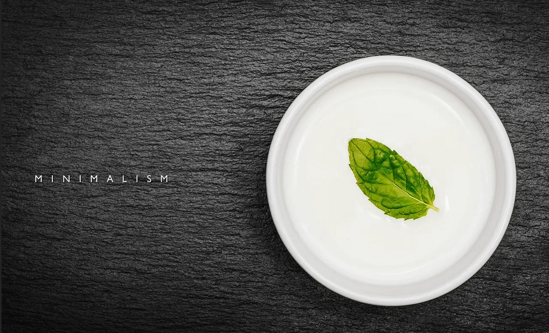
    Contemplation and respect for nature: Food as a form of meditation. It is necessary not to drown out the taste, but to hear it.

    Mindfulness in food: Every taste is felt, lived. No receptor overload.

    Balance: There is no single dominant taste in the dish, everything is balanced (sweet, sour, salty, bitter, umami).

    Simplicity = respect: If the product is good, why hide it behind sauces? Let him reveal himself.

    Minimalism of taste is not a rejection of taste, but an attempt to hear it on a deeper level. . It requires subtlety, habit and attention.

    What should be excluded:

    Flour. in all kinds and forms.
    Sugar in all kinds and forms. Including sugar-containing products, juices and waters.
    Hot spices.
    Any fast food and food in cafes and restaurants.
    Any refined products and semi-finished products.
    Any FRENCH fries.
    Any alcohol.
    Tobacco and drugs.

    and here’s why:

    Here’s how these foods and substances affect the body’s dopamine system::

    1. Flour (any kind)

    • Effect: A rapid increase in dopamine due to a sharp jump in blood glucose → a subsequent drop below normal.
    • Effects: A ‘splash-dip’ cycle is formed, which leads to dependence on flour products to raise your mood.

    2. Sugar (including hidden forms)

    • Mechanism: Sugar stimulates the release of dopamine in the nucleus accumbens (pleasure center) just like nicotine or cocaine.
    • Research: According to brain scans, sugar triggers a dopamine response comparable to drugs.
    • Risks: Reduced receptor sensitivity → more sugar is needed to produce the same effect.

    3. Hot spices (capsaicin)

    • Effect: Capsaicin triggers the release of dopamine through activation of TRPV1 receptors (the ‘euphoric’ effect after a spicy meal).
    • Feature: Unlike sugar, it does not cause a pronounced ‘kickback’, but it can form a psychological dependence.

    4. Fast food and restaurant food

    • Triggers: A combination of fat+salt+sugar → a powerful dopamine response (30% stronger than from home-cooked food).
    • Experiments: In an MRI scan, fast food activates the same areas as cocaine.
    • Especially dangerous: Monosodium glutamate (increases the ‘dopamine surge’ by 2-3 times).

    5. Refined products / semi-finished products

    • Problem: Artificially high concentration of ‘taste triggers’ (salt/sugar/fat) → depletion of dopamine receptors.
    • Effect: After consuming them, natural food seems ‘fresh’ (it takes time to restore sensitivity).

    6. Fried foods (especially fries)

    • Key Factor: Acrylamide (formed during frying) → increases dopamine by 40%, but is toxic to neurons.
    • Optional: The combination of crispy crust+fat creates a ‘perfect storm’ for the pleasure center.

    7. Alcohol

    • Action: Blocks GABA receptors → dopamine increases 5-10 times from normal.
    • Danger: Each dose of alcohol reduces the baseline dopamine level (more and more are needed to achieve the effect).

    8. Tobacco/Drugs

    • Nicotine: Increases dopamine by 200% in 10 seconds (the fastest delivery mechanism).
    • Heavy drugs: Can increase dopamine by 10-15 times, but completely destroy the natural system of its production.

    How to restore dopamine balance?

    1. Detox 21 days – complete rejection of the above (time to ‘reset’ the receptors).
    2. Natural Dopamine Stimulants:
    • Cold shower
    • Physical activity
    • Meditation
    1. Nutritional support:
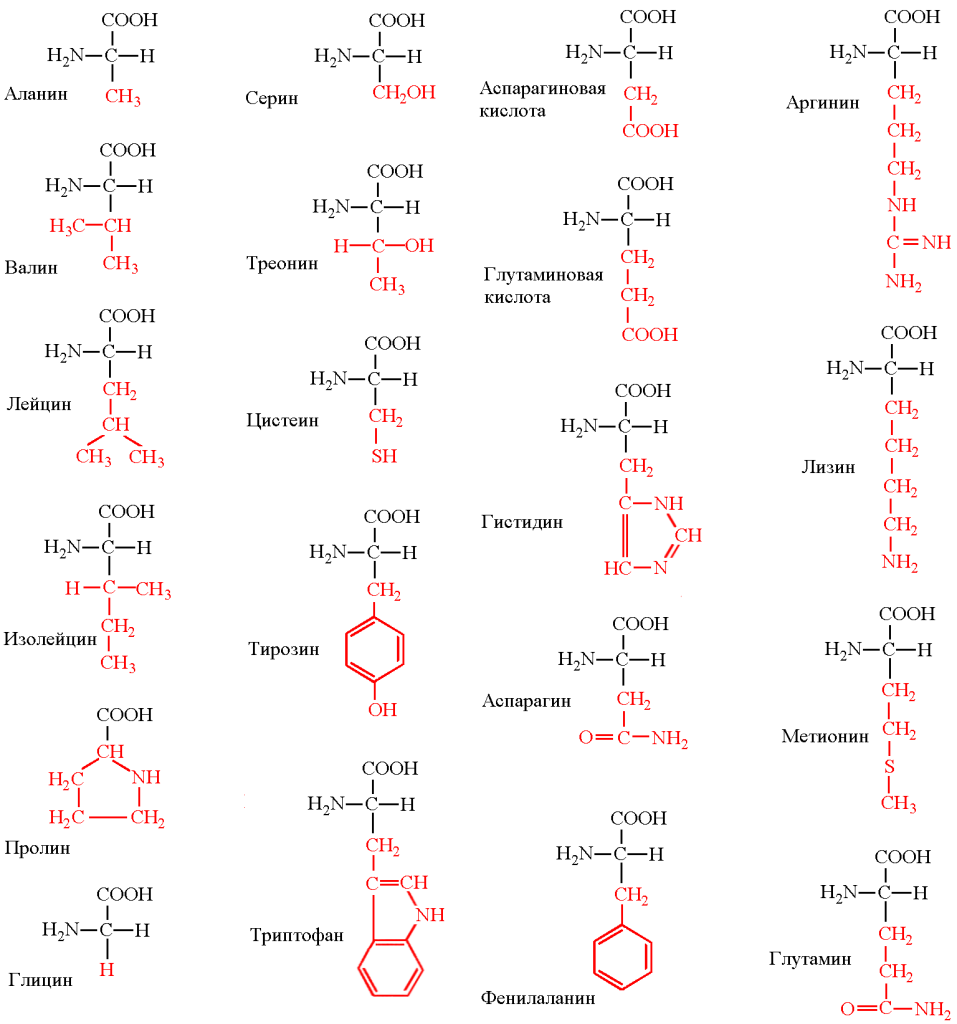
    • L-tyrosine (meat, fish, avocado)
    • Probiotics (fermented milk products)
    • Curcumin (suppresses inflammation in the dopamine pathways)

    Important: Full recovery of receptor sensitivity takes 3-12 months, depending on the’ experience ‘ of using the listed products.

    And then what happens? and here’s what:

    At first, stress and discomfort may occur, but then positive changes will follow.

    What will happen in the short term (first days-weeks)?

    • Breaking down sugar and fast carbs

    Irritability, fatigue, and headaches may appear-this is a reaction to the rejection of sugar and refined carbohydrates (as with a mild ‘detox’ reaction).

    Cravings for sweet and flour products will be strong, but after 1-2 weeks they will decrease.

    • Changes in digestion

    If there was a lot of flour in the diet before, temporary constipation is possible due to a decrease in the amount of fast carbohydrates.

    Bloating will decrease (especially if you used to eat a lot of yeast bread or sweet pastries).

    • Reduced caloric intake

    As you cut out high-calorie foods, your weight may start to drop (if there was an excess).

    What will happen in the long run (a month or more)?

    • Improved metabolism

    Reduce the risk of insulin resistance and type 2 diabetes.

    Blood sugar levels will return to normal, and sudden energy spikes and drowsiness after eating will disappear.

    • Weight loss (if you are overweight)

    The exclusion of flour and sugar dramatically reduces ’empty’ calories, which contributes to weight loss.

    • Improvement of skin condition

    Less sugar → less inflammation, acne, and premature aging (collagen glycation).

    It is possible to reduce swelling (if you used to eat a lot of hidden salt in fast food).

    • Energy Boost

    There will be no sudden spikes in glucose → more stable energy levels throughout the day.

    • Changing your taste habits

    Vegetables and natural products will start to taste better.

    Hot spices will not ‘overload’ the receptors, and you will begin to feel the natural tastes of food.

    • Improving the functioning of the gastrointestinal tract

    Less yeast, sugar, and refined foods → healthier gut microbiota.

    • Reducing the risk of inflammation

    Fast food and excess sugar provoke chronic inflammation, and avoiding them reduces this effect.

    Possible difficulties

    • Social issues – it is more difficult to eat out, and you may have questions from others.
    • Lack of certain vitamins (if you do not replace flour with healthy carbohydrates) – for example, B vitamins (in whole-grain bread).
    • Risk of disruptions – if you can’t find alternatives (for example, fruit instead of sweets, homemade food instead of fast food).

    Avoiding these products can significantly affect the search for meaning in life, but the mechanism of this influence is ambiguous. Here’s how it works:

    1. The connection between dopamine and existential Search

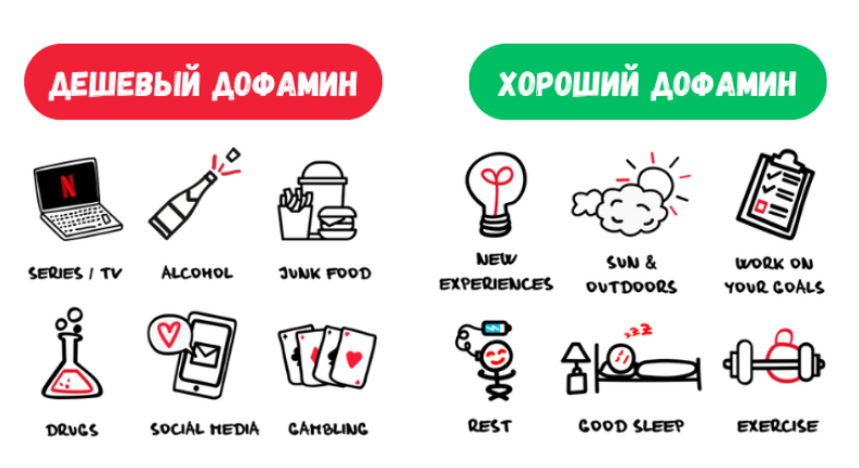
    • When chronically stimulated by ‘fast’ dopamine spikes (sugar, fast food, social media), the brain stops responding to natural sources of joy and meaning.
    • Studies show that people with an impaired dopamine system are more likely to experience an existential crisis (Journal of Neuroscience & Biobehavioral Reviews, 2021).

    2. What gives a refusal?

    • Restoring sensitivity to ‘slow dopamine’:
    • The joy of small achievements
    • Satisfaction from deep work
    • Interest in challenging tasks
    • Reduced anxiety (dopamine hyperstimulation increases the ‘I want more’ cycle → frustration)
    • Increased neuroplasticity – the brain’s ability to readjust and find new patterns of meaning

    3. Reverse side (first 2-4 weeks)

    • An existential void effect is possible:
    • The brain, devoid of the usual ‘crutches’, takes time to rebuild
    • The questions ‘What’s the point?’ may become more acute due to the lack of artificial incentives
    • This is a normal stage -a sign of the beginning of the transition to a more conscious search

    4. How to Increase your Positive Influence

    • Substitute practices:
    • Keeping a diary (structures thoughts)
    • Nature (phytoncides normalize dopamine)
    • Social interactions without stimulants (live conversations instead of ‘dopamine’ messages)
    • Philosophical work:
    • Reading the existentialists (Camus, Frankl) against the background of a purified perception gives other insights
    • Practice of ‘ death meditation ‘( representing the extremity of life)

    Example from research:
    In the MIT experiment (2023) participants after 6 weeks of abstaining from hyperstimulating food:

    • 37% more likely to report a ‘sense of meaning’ in everyday activities
    • 28% less likely to ask the question ‘ Why all this?’

    Conclusion:

    A refusal won’t give you a complete answer about the meaning of life, but:
    ✅ It will create neurobiological conditions for its search
    ✅ It will remove the ‘noise’ that prevents you from hearing yourself
    . It will restore the ability to find meaning in small things

    It’s like taking off your glasses with dirty lenses – the world won’t change, but you’ll start to see it more clearly. The main thing is to survive the transition period (usually 21-40 days).

  • μ- (mu) and δ — (delta) receptors

    μ- (mu) and δ — (delta) receptors

    mk- (mu) and δ — (delta) receptors are the two main types of opioid receptors in the human body that are part of the complex endogenous opioid system. This system plays a key role in regulating pain, reward, and many other physiological processes.

    Both receptors belong to the class of G-protein coupled receptors (GPCRs), and their activation leads mainly to inhibitory effects in the nervous system.

    For clarity, here is a comparative table of their main characteristics:

    Characteristicμ-receptor (Mu receptor)δ-receptor (Delta receptor)
    Primary functionsAnalgesia (pain relief)
    Euphoria, sense of well-being
    Respiratory depression
    Constipation
    Physical dependence
    • Sedative effect
    Analgesia (especially in chronic pain)
    Mood regulation (antidepressant effect)
    Neuroprotection
    • Modulation of μ-receptor-mediated effects
    Endogenous ligands
    (natural body agents)
    β-endorphin (high affinity)
    Enkephalins (moderate affinity)
    Enkephalins (high affinity)
    β-endorphin (moderate affinity)
    Exogenous agonists
    (external activators)
    Morphine, heroin, fentanyl,
    methadone, oxycodone
    Leu-enkephalin, Met-enkephalin (unstable)
    SNC80 (experimental compound)
    Antagonists
    (blockers)
    Naloxone, naltrexoneNaltrindole (experimental)
    Key effects upon activationPotent analgesia, euphoria, respiratory depression (main cause of death in overdose), miosis (pupil constriction), reduced gastrointestinal motility.Analgesia (less potent but with lower risk of respiratory depression), antidepressant effect, modulation of μ-receptor-mediated responses.
    CNS localizationHigh density in:
    Thalamus (pain relay center)
    Periaqueductal gray matter (PAG)
    Brainstem (respiratory center)
    More restricted distribution:
    Olfactory bulbs
    Neocortex
    Limbic system (emotion-related)
    Addiction potentialVery high. Primary mediator of the rewarding effects (pleasure/reinforcement) of opioids.Low. δ-receptor activation alone does not produce significant euphoria or physical dependence.

    Learn more about the mk-receptor (Mu-receptor)

    This is the most well-known and clinically significant receptor. It is associated with both the powerful therapeutic effects of opioid analgesics and their main dangerous side effects.

    • Role in medicine: It is the main target for morphine and other powerful painkillers used to relieve acute and severe chronic pain (for example, in cancer).
    • Side effects:
      • Respiratory depression: The most dangerous effect. mk-agonists reduce the sensitivity of the respiratory center in the brain stem to CO₂. If you overdose, your breathing simply stops.
      • Constipation: Activation of m-receptors in the gut drastically reduces intestinal motility, which is a common problem for patients taking long-term opioids.
      • Euphoria and addiction: A strong sense of pleasure and well-being caused by the activation of m-receptors in the brain’s mesolimbic pathway is a major cause of opioid drug addiction and abuse.

    Learn more about the Delta receptor

    The role of δ-receptors is less well understood, but no less important. They often work in tandem with m-receptors.

    • Prospects for pharmacology: Scientists are actively looking for delta-receptor agonists, as there is evidence that they can provide pain relief with a lower risk of respiratory depression and addiction development. . This could be a breakthrough in creating safe analgesics.
    • Influence on mood: Activation of δ-receptors demonstrates antidepressant and anxiolytic (anti-anxiety) effects in preclinical studies.
    • Modulation: It is believed that δ-receptors modulate the activity of μ-receptors. For example, blockade of δ-receptors may enhance some of the effects of μ-agonists.

    Interaction and meaning

    Opioid receptors rarely work in isolation. Their interaction creates a complex regulatory network. For example, some drugs aim to be bifunctional agonists (for example, activate both m-and d-receptors) or agonist-antagonists (block one type and activate the other) to try to separate desirable effects (pain relief) from undesirable ones (respiratory depression, addiction).

  • Follistatin plays an important role in regulating cell growth and differentiation

    Follistatin plays an important role in regulating cell growth and differentiation

    Follistatin. is a protein that plays an important role in regulating cell growth and differentiation, especially in the context of muscle tissue and the reproductive system. It is known as an antagonist of myostatin , a protein that restricts muscle growth. Follistatin binds and inhibits myostatin, as well as other members of the TGF-β (Transforming Growth factor beta) family, making it a key regulator of muscle growth and tissue repair.


    Main functions of follistatin:

    1. Inhibition of myostatin:
      • Myostatin is a protein that inhibits the growth and differentiation of muscle cells. Follistatin binds myostatin, preventing its interaction with receptors, which leads to increased muscle growth.
      • This property makes follistatin promising for the treatment of diseases associated with muscle atrophy, such as muscular dystrophy.
    2. Regulation of the TGF-β signaling pathway:
      • Follistatin also binds and inhibits other members of the TGF-β family, such as activin and BMP (Bone Morphogenetic protein). These proteins are involved in the regulation of cell growth, differentiation, and apoptosis (programmed cell death).
    3. Role in the reproductive system:
      • Follistatin is produced in the ovaries and testicles, where it regulates the processes of folliculogenesis (development of follicles in the ovaries) and spermatogenesis (sperm formation).
      • It is also involved in regulating the level of follicle-stimulating hormone (FSH), which is important for reproductive function.
    4. Participation in tissue repair:
      • Follistatin promotes tissue regeneration, including muscle, bone, and skin, due to its ability to modulate the TGF-β signaling pathway.

    Application of follistatin:

    1. Medicine:
      • Treatment of muscle diseases: Follistatin is being studied as a potential treatment for muscular dystrophy and other diseases associated with muscle atrophy.
      • Regenerative Medicine: Its ability to stimulate tissue growth makes it promising for recovery from injuries and surgeries.
      • Reproductive Medicine: Follistatin can be used to treat fertility disorders.
    2. Sport:
      • Due to its ability to boost muscle growth, follistatin has attracted attention in sports medicine. However, its use in sports can be considered doping and is prohibited by anti-doping agencies.
    3. Aging research:
      • Follistatin is being studied in the context of slowing age-related muscle loss (sarcopenia) and improving fitness in older adults.

    Mechanism of action:

    Follistatin acts by binding to myostatin, activin, and other members of the TGF-β family. This prevents them from interacting with receptors on the cell surface, which blocks signaling pathways responsible for suppressing muscle and other tissue growth.


    Researches:

    • Animal studies have shown that increasing follistatin levels leads to a significant increase in muscle mass and strength.
    • In human clinical trials, follistatin is being studied for the treatment of muscular dystrophy and other diseases. However, its long-term safety and efficacy are not yet fully understood.

    Conclusion:

    Follistatin is a multifunctional protein that plays a key role in regulating muscle growth, reproductive function, and tissue repair. Its ability to inhibit myostatin makes it promising for treating muscle diseases and improving fitness. However, further research is needed to fully understand its potential and safety for clinical use.

  • Amino acids-what they consist of, classification, role

    Amino acids-what they consist of, classification, role

    Amino acids are organic compounds that are the main building blocks of proteins. They play a key role in biological processes such as protein synthesis, cell signaling, and metabolism. Each amino acid consists of certain chemical elements and functional groups. Let’s analyze their structure in detail.


    Main components of the amino acid:

    1. Central carbon atom (α-carbon):
      • This is the central atom in the structure of an amino acid, to which all other groups are attached.
      • It is a chiral center (with the exception of glycine), which means that amino acids can exist as two optical isomers (L — and D-forms). In nature, L-amino acids are more common.
    2. Amino Group (-NH₂):
      • This is the main group, which contains a nitrogen atom bound to two hydrogen atoms.
      • The amino group gives the amino acid its basic properties.
    3. Carboxyl group (-COOH):
      • It is an acidic group consisting of a carbon atom bonded to two oxygen atoms and one hydrogen atom.
      • The carboxyl group gives the amino acid acidic properties.
    4. Hydrogen Atom (-H):
      • A simple hydrogen atom bound to an α-carbon.
    5. R-group (side chain):
      • It is a unique part of an amino acid that distinguishes one amino acid from another.
      • The R-group can be simple (like glycine, where it is just a hydrogen atom) or complex (like arginine, where it is a long carbon chain with additional functional groups).
      • The properties of the R-group determine the chemical and physical characteristics of the amino acid (for example, hydrophobicity, hydrophilicity, charge).

    General Amino Acid Formula:

    • H is hydrogen.
    • nh₂ is an amino group.
    • COOH is a carboxyl group.
    • R — side chain (variable part).

    Classification of amino acids:

    Amino acids are classified according to the properties of their R-groups:

    1. Nonpolar (hydrophobic):
      • Examples: alanine, valine, leucine, isoleucine.
      • These amino acids are poorly soluble in water.
    2. Polar uncharged:
      • Examples: serine, threonine, asparagine, glutamine.
      • These amino acids have polar side chains, but do not carry a charge.
    3. Charged (acidic and basic):
      • Acidic: aspartic acid, glutamic acid (have a negative charge).
      • Basic: lysine, arginine, histidine (have a positive charge).
    4. Aromatic products:
      • Examples: phenylalanine, tyrosine, tryptophan.
      • These amino acids contain aromatic rings in their side chains.

    The role of amino acids in the body:

    1. Protein Synthesis: Amino acids are linked in chains through peptide bonds to form proteins.
    2. Metabolism: Some amino acids are involved in the synthesis of other important molecules, such as neurotransmitters (eg, glutamate, GABA).
    3. Energy metabolism: Amino acids can be used as a source of energy during digestion.
    4. Signaling: Some amino acids (such as glycine, glutamate) function as neurotransmitters.

    Conclusion

    Amino acids are fundamental molecules consisting of an amino group, a carboxyl group, hydrogen, and a unique side chain (R-group). Their diversity and properties determine their role in protein construction and other biological processes. Understanding the structure of amino acids helps to understand their functions and significance for life.

  • Dopamine addiction

    Dopamine addiction

    Dopamine addiction is a complex condition that can include cravings for sweets, alcohol, drugs, gambling, social media, and other sources of quick pleasure. Different approaches can be used to combat this addiction, including medication, psychotherapy, and behavioral methods.

    1. Drugs that reduce dopamine cravings

    Some medications can help reduce excessive dopamine system activity and reduce addiction:

    A) To combat food addiction (sweet, overeating)

    • Naltrexone + bupropion (Contrave / Mizar) – reduces food cravings and blocks the pleasure of overeating.
    • Liraglutide (Saxenda, Victosa– is a GLP-1 agonist that reduces appetite and cravings for high-calorie foods.
    • Topiramate– Topamax) — sometimes used off-label to reduce cravings for food and alcohol.

    B) For alcohol and drug addiction

    • Naltrexone -blocks opioid receptors, reducing the enjoyment of alcohol and opiates.
    • Acamprosate (Campral) – normalizes the balance of GABA and glutamate, reducing cravings for alcohol.
    • Disulfiram (Antabuse) — causes alcohol poisoning by forming negative reinforcement.
    • Bupropion (Wellbutrin, Zyban) — can reduce cravings for nicotine and overeating.

    C) For behavioral addictions (gambling, social networks, shopping)

    • Naltrexone -sometimes used to reduce impulsivity.
    • SSRIs (fluoxetine, sertraline, escitalopram – may reduce compulsive behavior.
    • Atomoxetine (Strattera) — a noradrenaline drug that improves pulse control.

    2. Non-medicinal methods

    • Cognitive Behavioral therapy (CBT) – helps you rebuild your thinking and response to triggers.
    • Meditation and mindfulness -reduce impulsivity and increase self-control.
    • Exercise is a natural way to boost dopamine and serotonin without harm.
    • Dopamine detox is a temporary withdrawal from hyperstimulating activities.

    3. Natural ways to regulate dopamine

    • L-tyrosine / phenylalanine are dopamine precursors.
    • Magnesium and Omega-3 support the balance of neurotransmitters.
    • Adaptogens (Rhodiola rosea, ginkgo biloba) — help reduce stress and normalize dopamine.

    Conclusion

    The best approach is a combination of medications (if prescribed by a doctor), therapy, and lifestyle changes. If the addiction is severe (drugs, alcoholism), it is important to consult a narcologist or psychiatrist. For light forms (food, social networks), behavioral methods and working with a psychologist are often enough.

  • Glandular epithelium

    Glandular epithelium

    Glandular epithelium is a type of epithelial tissue that specializes in the secretion of various substances. It forms glands that can be either part of organs or independent structures. The glandular epithelium is involved in the production and release of enzymes, hormones, mucus, sweat, saliva and other biologically active substances necessary for the functioning of the body.

    Main characteristics of glandular epithelium:

    1. Cellular structure: Glandular epithelial cells are usually cubic or cylindrical in shape and contain well-developed organelles, such as the endoplasmic reticulum and Golgi apparatus, which are essential for the synthesis and secretion of substances.
    2. Secretory function: Cells can secrete substances in various ways:
      • Merocrine type: The secret is released without destroying the cell (for example, salivary glands).
      • Apocrine type: Part of the cell is rejected along with the secret (for example, mammary glands).
      • Holocrine type: The cell is completely destroyed, releasing a secret (for example, sebaceous glands).

    Types of glands:

    1. Exocrine glands: Secrete a secret on the surface of the body or in the cavities of organs through ducts. Examples:
      • Sweat glands (secrete sweat).
      • Salivary glands (secrete saliva).
      • The pancreas (secretes digestive enzymes).
    2. Endocrine glands: Secrete hormones directly into the blood or lymph, without having ducts. Examples:
      • The thyroid gland (secretes thyroxine).
      • The adrenal glands (which secrete epinephrine and cortisol).
      • Pituitary gland (secretes hormones that regulate other glands).

    Glandular epithelial functions:

    • Regulation of metabolism: Hormones secreted by the endocrine glands regulate metabolism, growth, and development.
    • Digestion: Enzymes secreted by the exocrine glands are involved in the digestion of food.
    • Protection: Mucus secreted by the glands protects the mucous membranes from damage and infection.
    • Thermoregulation: The sweat glands help cool the body.

    Glandular epithelium plays an important role in maintaining homeostasis and adapting the body to changing environmental conditions.